Research
Coherence Domain Probing Systems
The development of various endoscopic OCT probes
has greatly extended the application range of this high-resolution
biomedical imaging technique. One of our research interest is
a new design for a forward-imaging OCT needle probe – the
Paired Angle Rotation Scanning OCT (PARS-OCT) probe. Based on
our PARS technology, we developed the hand-held forward-imaging
optical coherence tomography (OCT) needle endoscope system. The
feasibility of retinal imaging has been tested on enucleated
ex vivo porcine eyes, where structural features including remnant
vitreous humor, retina, and choroid can be clearly distinguished.
The narrow probe can easily fit through the standard cannula
or scleral incisions employed in ophthalmic surgery. The probe
can potentially serve as a better alternative to traditional
visual inspection by white-light illumination during vitreoretinal
surgery (e.g. vitrectomy).
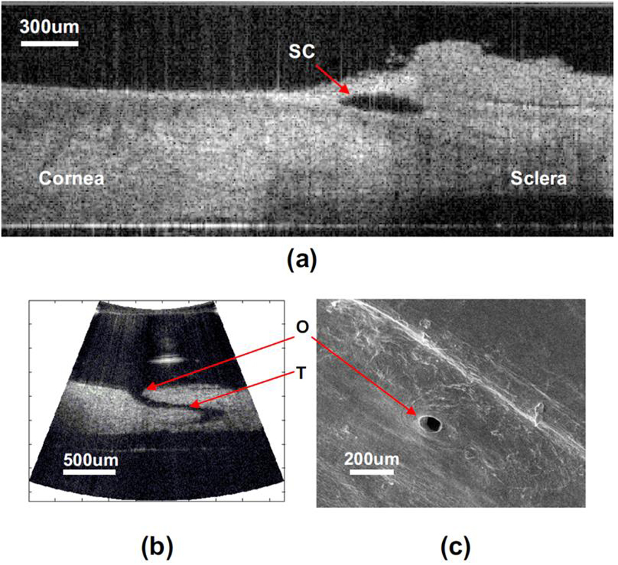
The figure above illustrates OCT and SEM images of human cadaver eye tissue segments:
(a) OCT image of a tissue segment with TM intact, acquired by
translating the stage. (b) OCT image of a tissue with TM removed,
acquired by rotating the needles of the endoscopic probe. (c) SEM
image of the same CC in (b). O, CC opening; T, CC path through sclera.

|