Fourier Ptychography
The standard microscope is a time tested and vital piece of equipment in a life science lab. Yet, its performance has undergone only gradual improvements over time. This is because physical optical lenses are intrinsically imperfect. Building better microscopes has traditionally focused on using ever more complicated optical element arrangements to minimize aberrations. Fourier Ptychography is a computational microscopy method that shifts the challenge of addressing aberrations from the physical world to the computational realm where aberrations become readily solvable mathematical problems. In the process, we enable a standard microscope to push past its physical optical limitations to provide digital refocusing, improved spatial-bandwidth product and phase imaging capabilities.
Optical Setup:
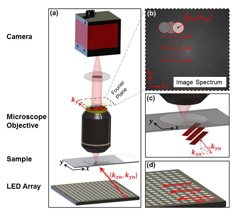
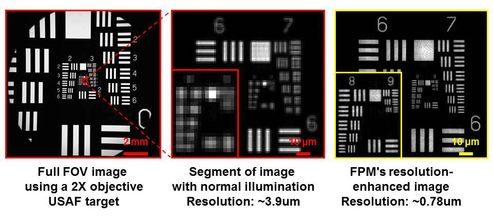
Demonstration of resolution enhancement
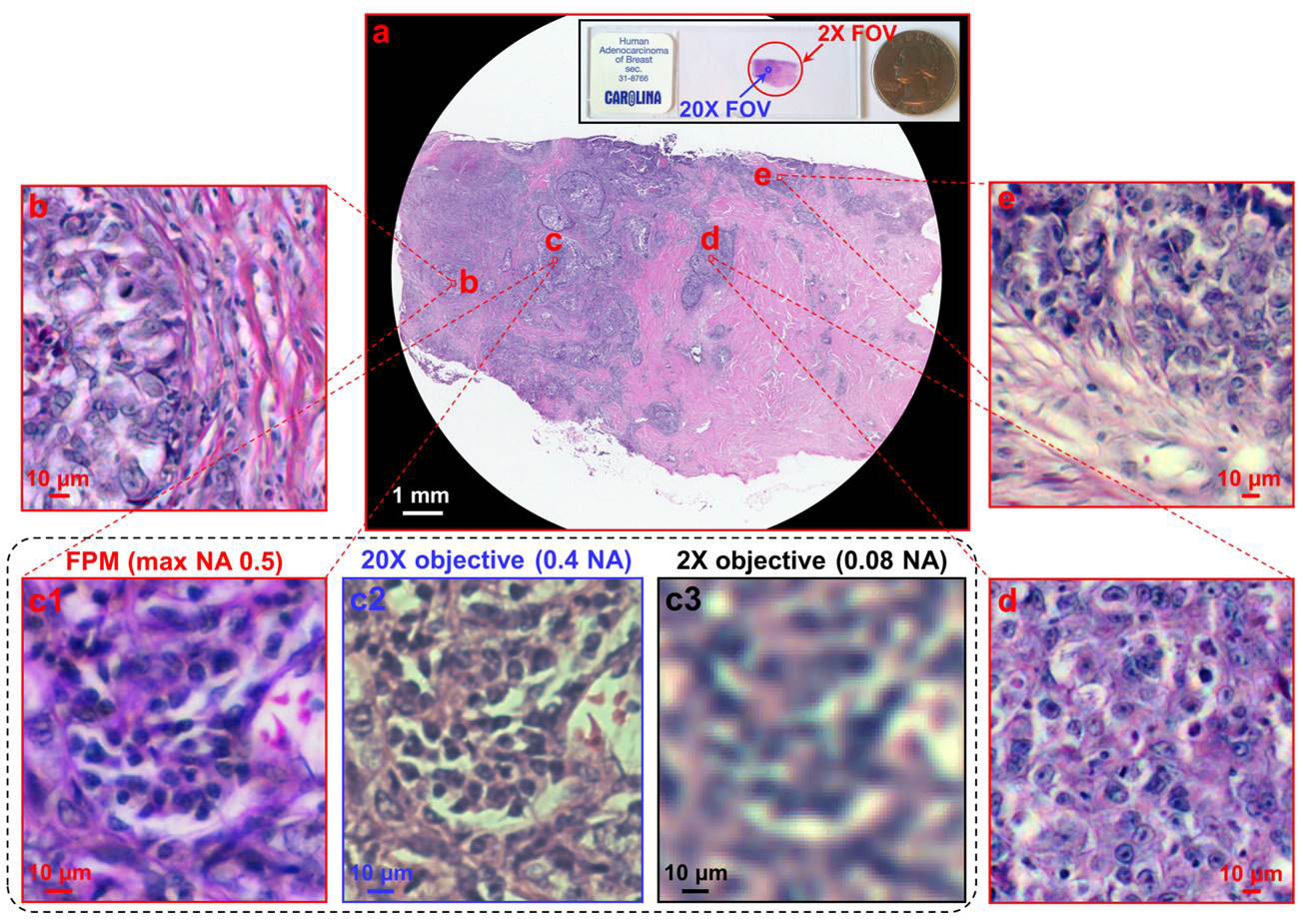
Imaging biological samples with FPM
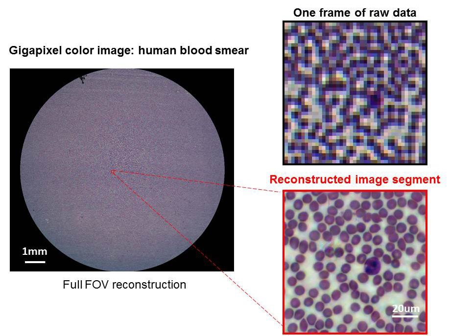
Digital Pathology
The figure above shows a gigapixel color imaging of a pathology slide. Insets demonstrate detail with comparison to a conventional microscope image
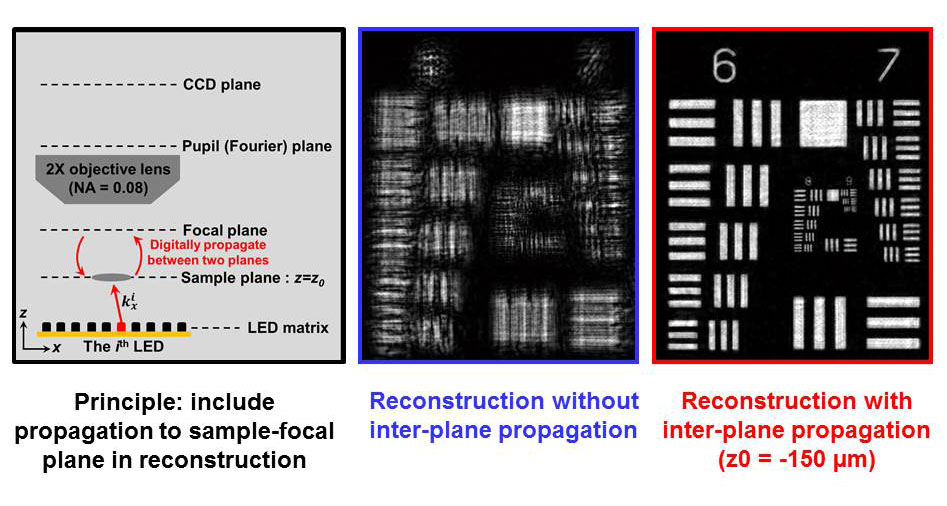
Extending image depth-of-focus with digital refocusing
- FPM digitally corrects for slide positioning and tilt errors
Embedded pupil function recovery (EPRY)
For any lens, its ability to form a sharp image degrades as you move away from its optical axis. This is because the lenss
spherical surfaces begin to fail to approximate the properties of an ideal lens and introduce aberrations. With a small modification,
the Fourier ptychographic reconstruction algorithm can not only reconstruct a high spatial-bandwidth, complex field of the sample, but
also quantify the spatially varying aberration across the microscopes entire field of view in the form of pupil functions [3]. As a result,
we can obtain an aberration-free image over the entire FOV and also the systematic aberration, which can be a valuable piece of
information for calibrating the imaging system.
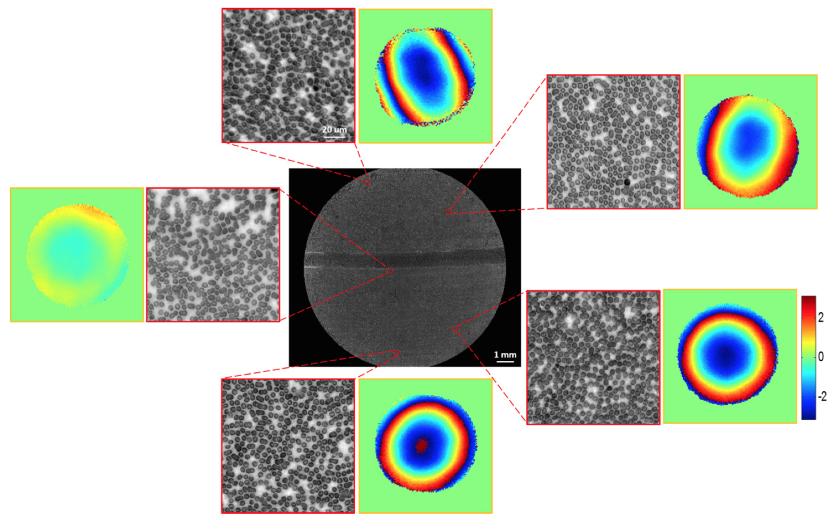
The figure above shows the high resolution image of a blood smear in different regions of the FOV along with the pupil functions, which clearly show their spatially-varying nature.
Wide FOV aberration-free fluorescence imaging
Fluorescence imaging is used extensively by biologists to study cells functions and behaviors. A standard microscope, fitted with excitation and emission filters, is typically used to excite and collect signals from specific fluorophores in a sample. Thus, a fluorescence image captured with the microscope is under the influence of the same aberrations as a bright-field image. We demonstrate that we can first acquire an FPM dataset, with the LED array as the light source, to reconstruct both the sample’s complex field and the spatially-varying pupil functions. We can then use the pupil functions to remove aberrations in the fluorescence image captured with a standard fluorescence imaging method.
The result is a set of wide FOV, aberration-free, and multimodal microscopic images that contains both the morphological and fluorescence information [4].
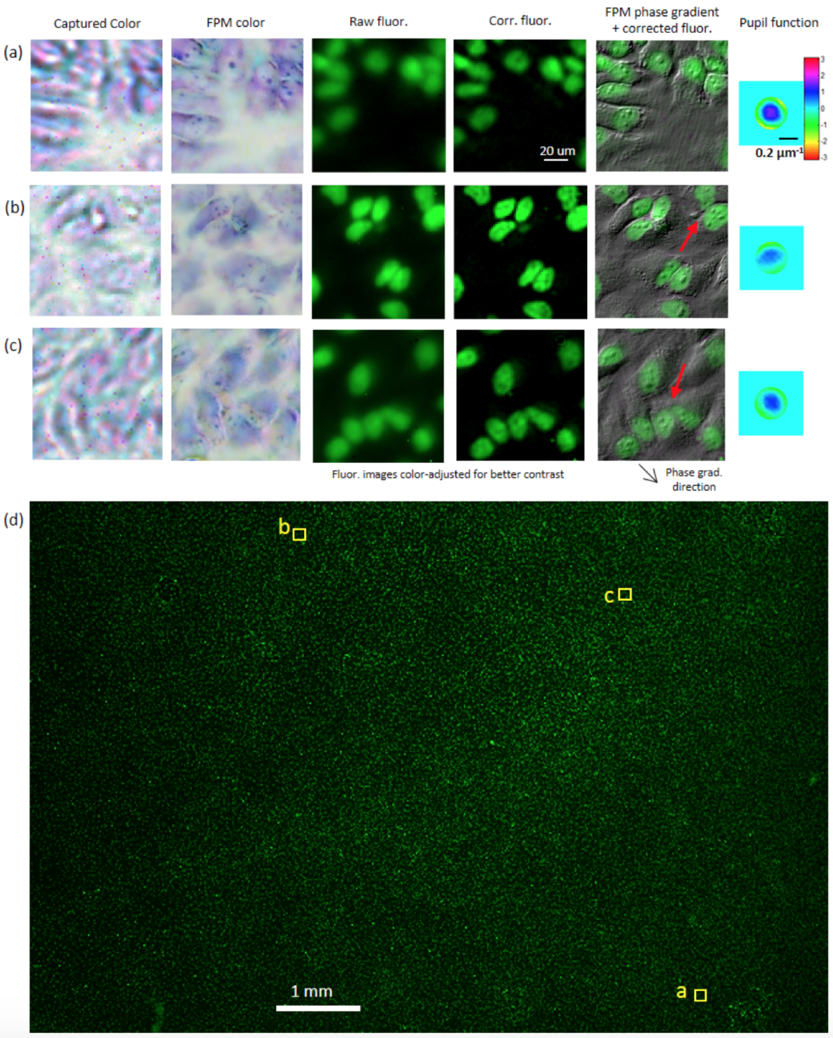
The figure above shows a sample of HeLa cells fluorescently tagged by DAPI (color adjusted to green for visualization). Images combining the fluorescence signal and the FPM-reconstructed phase are rich in information.

References
[1] Guoan Zheng, Roarke Horstmeyer and Changhuei Yang, "Wide-field, high-resolution Fourier ptychographic microscopy," Nature Photonics, doi:10.1038/nphoton.2013.187
[2] Xiaoze Ou, Roarke Horstmeyer, Changhuei Yang, and Guoan Zheng, "Quantitative phase imaging via Fourier ptychographic microscopy (FPM)," submitted.
[3] Xiaoze Ou, Guoan Zheng and Changhuei Yang, "Embedded pupil function recovery for Fourier ptychographic microscopy," Optics Express 22 4960-72 (2014)
[4] Jaebum Chung, Jinho Kim, Xiaoze Ou, Roarke Horstmeyer and Changhuei Yang, Wide field-of-view fluorescence image deconvolution with aberration-estimation from Fourier ptychography, Biomedical Optics Express 7 352-368 (2016)
|