Research
PARS-OCT Forward Imaging Probe
Over the past decade, the development of various endoscopic
OCT probes has greatly extended the application range of this
high-resolution biomedical imaging technique. One of our research
interest is a new design for a forward-imaging OCT needle probe – the
Paired Angle Rotation Scanning OCT (PARS-OCT) probe. This probe
design utilizes a pair of angle-cut rotating GRIN lenses to deflect
and scan the OCT probe beam across the forward region ahead of
the probe tip. In this design, the scan actuation system may
be located away from the probe tip, much like in the case of
a side-imaging OCT probe, enabling easy miniaturization of the
actual probe. Further, this probe design can achieve a large
forward scan arc length to probe diameter ratio. This parameter
is especially relevant for clinical probe considerations, as
a clinician will desire as wide a scan range as possible with
the smallest possible probe size.
The PARS-OCT probe channels the input OCT probe light from a
single mode fiber through the first GRIN lens [see the following
figure (a) and (b)]. The light beam exits from the other face
of the GRIN lens which is cut at an angle Ψ. The beam then
enters the second GRIN lens through an identically angle-cut
face of the GRIN lens. Finally, the beam exits the second GRIN
lens and focuses at a point ahead of the probe. The exact focal
point is determined by the pitch of the two GRIN lenses.
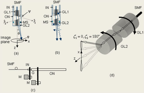
We define the orientations of the two GRIN lenses
by angles ζ1 and ζ 2, which are defined as the angles
between the projections of vectors
 and and
 , respectively,
in the image plane and the x-axis [see figure (a)]. An analytical expression of
the angle θ as a function of ζ1, ζ2 (under small
angle approximation) can be derived as: , respectively,
in the image plane and the x-axis [see figure (a)]. An analytical expression of
the angle θ as a function of ζ1, ζ2 (under small
angle approximation) can be derived as:
(1)

where
(2)
and  are the on-axis refractive index and the index gradient constant
of the GRIN lens, respectively. Z is the length of the second
GRIN lens and d is the diameter of the GRIN lens. are the on-axis refractive index and the index gradient constant
of the GRIN lens, respectively. Z is the length of the second
GRIN lens and d is the diameter of the GRIN lens.
A fan sweep of the output beam in xz-plane [shown vertical in
figure (d)] can be performed by simply rotating the two GRIN
lenses in opposite directions at the same angular speed.
The PARS-OCT probe design is capable of performing volumetric
scans with very little modifications. By simply incrementally
off-shifting the relative orientation of the two GRIN lenses
while performing B-scans, we can acquire volumetric scans. A
simpler implementation will be to introduce a slight offset to
the relative rotation scan velocities. In this case, the acquired
B-scans will automatically sweep through the entire volume scan
space. Our acquisition of orthogonal B-scans with the prototype
probe demonstrates the simplicity by which volumetric scans may
be performed.
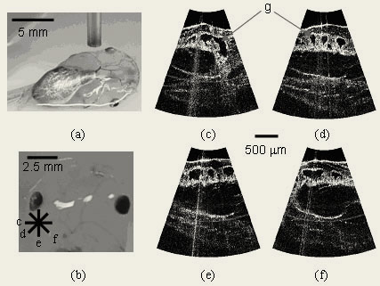
In the demonstration, we rotated the two needles with equal
and opposite angular speed (~21 rpm), and acquired a single B-scan
image from the specimen. We then rotated both needles by 45º increment
and acquired the second, third, and fourth B-scan image [see
the following figures (b)]. Fig. (a) shows the photograph of
the needle and the tadpole when acquiring the images. The scanned
locations are shown in Fig. (b). The acquired images are displayed
in Fig. (c)-(f). Each image has 350 A scan lines and is acquired
in 1.4 s. We can clearly discern the gill pockets in the images.
The scan depth in the image is 2.3 mm and the largest scan half-angle
is 19º.
The probe can be potentially used in needle guidance or biopsy
to provide high-resolution 3-D tomographic images of the targets
forward of the probe.

References
Jigang Wu, Michael Conry, Chunhui Gu, Fei Wang, Zahid Yaqoob,
and Changhuei Yang. “Paired-angle-rotation scanning optical
coherence tomography forward-imaging probe,” Optics Letters,
31, 1265, (2006). (pdf)
|