Research
Wide Field-of-view on-chip Fluorescence Microscopy
Because of the high specificity and high sensitivity of fluorescent probes, fluorescence microscopy plays a vital role in modern clinical diagnosis and biological research. However, due to its limited field-of-view (FOV), size, and cost, conventional microscopy is becoming a bottleneck in rapidly emerging and evolving areas such as large-scale genome screening, point-of-care diagnosis, and long-term cell imaging.
We have developed a wide FOV on-chip fluorescence microscopy method based on the Talbot effect, termed fluorescence Talbot microscopy (FTM) 1, 2 (Fig. 1-2). Unlike conventional fluorescence microscopy, the FOV for FTM can be arbitrarily scaled up without incurring a proportionate imaging time increase or sacrifice of resolution. Our FTM prototype has a resolution of 1.2 µm and an FOV of 3.9 mm × 3.5 mm.
We have demonstrated the capability of our system by imaging human breast cancer cells (SK-BR-3) stained by Qdot, live HeLa cells and C-elegans expressing GFP (Fig. 3-4). In addition, we were able to perform time-lapse imaging on live HeLa cells, and track the migration and division of single cells over an area of 13 mm2 (Fig. 5).
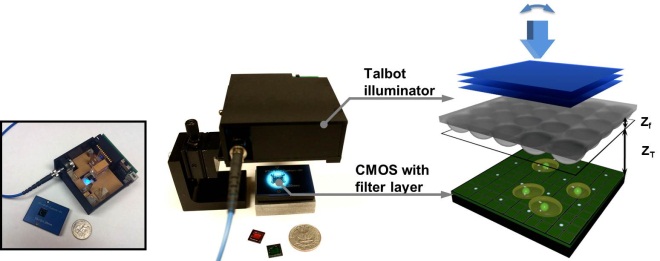
Fig. 1 FTM prototype
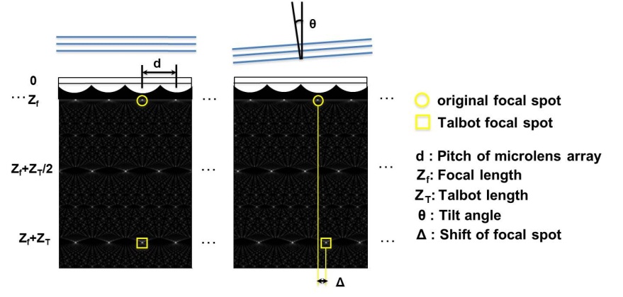
Fig. 2 FTM scanning principle
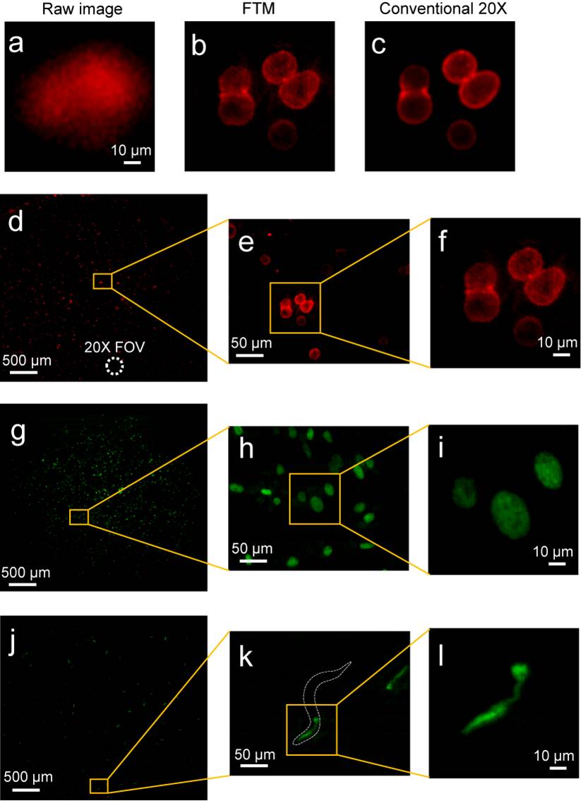
Fig. 3 (a-f) Fluorescence images of human breast cancer cells (SK-BR-3) with membranes stained by Qdot® 625. (d) The full FOV FTM image. (e, f) The magnified FTM image. (g-i) Fluorescence image of HeLa cells with GFP expression in nuclei. (j-l) Fluorescence image of C. elegans with GFP expression in pharynx.
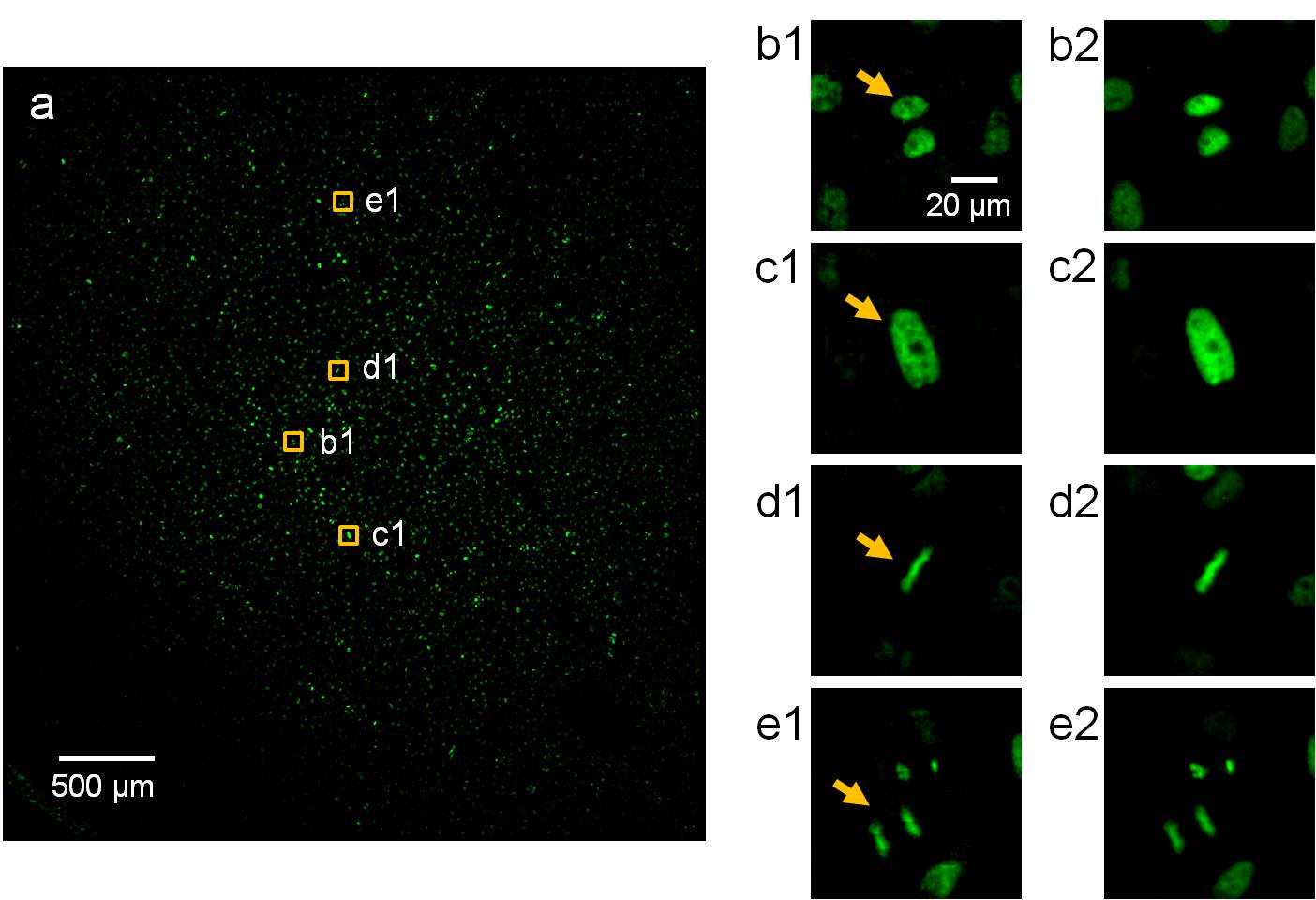
Fig. 4 FTM images of HeLa cells in different stages of the cell cycle. (a) The full FOV FTM image. (b1, c1, d1, e1) Cropped images of typical cells in (a), including G1 (b1), G2 (c1), metaphase (d1), and anaphase (e1) (arrows). (b2, c2, d2, e2) The same cells as imaged by a conventional microscope with a 20x/0.4 NA objective.
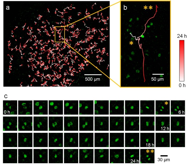
Fig. 5 FTM Time-lapse imaging of HeLa cells expressing GFP in nuclei. (a) Trajectories of cells over 24.8 hours. (b) The trajectory showing a single cell which divided at 5.5 h (*), with one of the daughter cells that moved upwards dividing again at 24.8 h (**). (c) Longitudinal images of the cell in (b) as it migrated and divided.
Wide Field-of-view Multicolor Fluorescence Microscopy
We further developed a multicolor fluorescence Talbot microscopy (multicolor FTM) system that is able to perform multicolor fluorescence microscopy imaging3 (Fig. 6). Unlike our on-chip Talbot microscopy implementation1,2 this system is capable of imaging standard microscopical slides over an FOV of 12 × 10 mm2 with an acquisition time as fast as 23 seconds for one fluorescence channel. As a demonstration, we imaged biological samples such as double-stained human breast cancer SK-BR-3 cells (Fig. 7), Giardia lamblia cysts, and the Cryptosporidium parvum oocysts (Fig. 8).
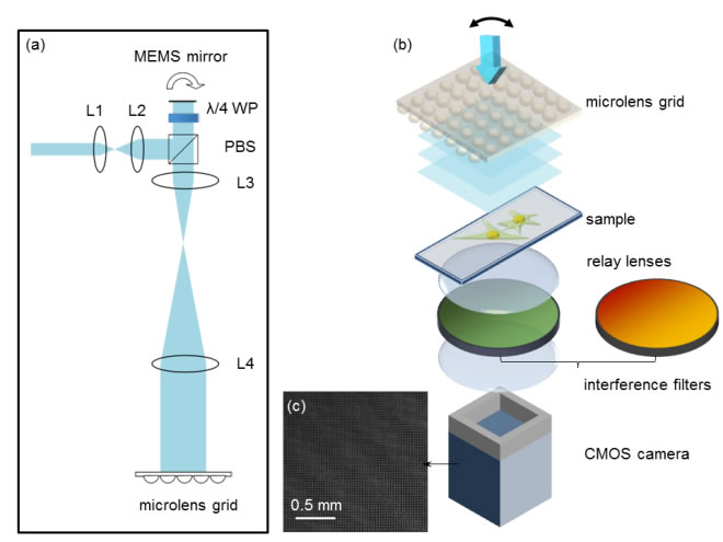
Fig. 6 The system setup for the Talbot microscope. (a) The optical setup for generating an angular shift incident beam to the microlens grid. (b) The system setup for the sample and detection. (c) The Talbot-focused spots.
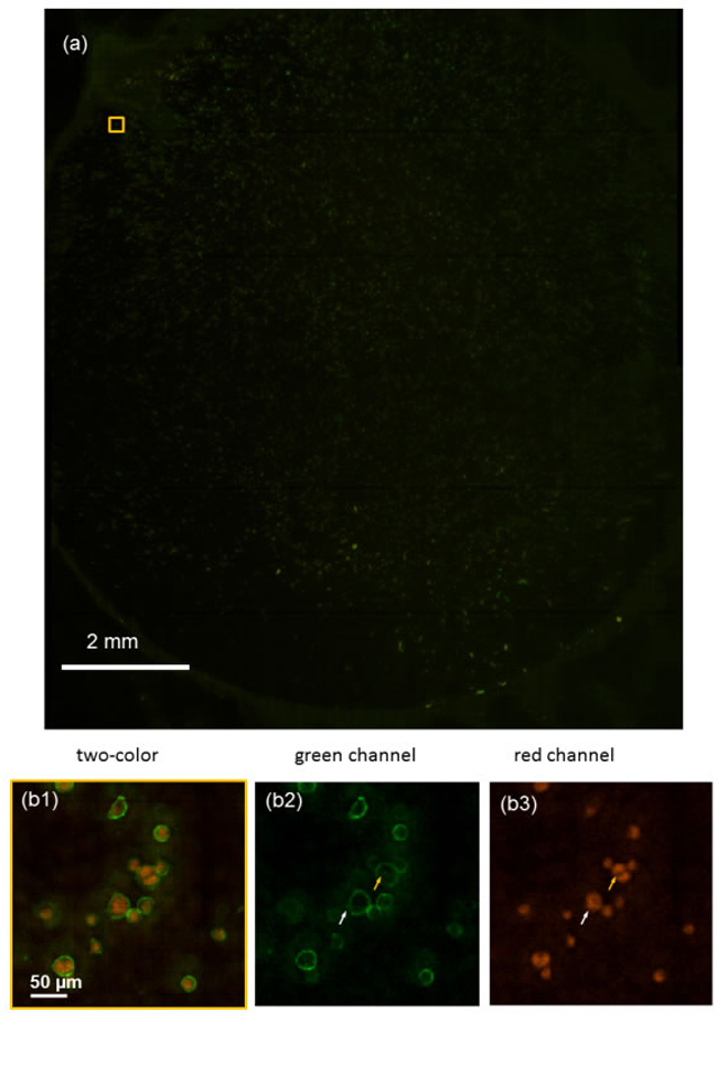
Fig. 7 Wide FOV two-color fluorescent images of human breast cancer SK-BR-3 cells.
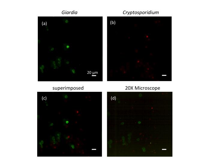
Fig. 8 The fluorescent image of a mixture of Giardia lamblia cysts labeled with Alexa Fluor® 488 and Cryptosporidum parvum oocysts labeled with Qdot® 625.

References
[1] Shuo Pang, Chao Han, Mihoko Kato, Paul W. Sternberg, and Changhuei Yang; "Wide and scalable field-of-view Talbot-grid-based fluorescence microscopy;" Optics Letters 37 (23): 5018-5020 (2012)
[2] Chao Han, Shuo Pang, Danielle V. Bower, Patrick Yiu, and Changhuei Yang; "Wide Field-of-View On-Chip Talbot Fluorescence Microscopy for Longitudinal Cell Culture Monitoring from within the Incubator;" Analytical Chemistry 85 (4): 2356-2360 (2013)
[3] Shuo Pang, Chao Han, Jessey Erath, Ana Rodriguez and Changhuei Yang; "Wide field-of-view Talbot grid-based microscopy for multicolor fluorescence imaging;" Optics Express 21 (12): 14555 (2013)
|