Research
Hand-Held Forward-Imaging Needle Endoscope for Ophthalmic
OCT Inspection
One potential application of our PARS probe is aiding vitrectomy,
which is a surgical procedure to remove the vitreous humor (sketched in Fig. 1).
Vitrectomy is a required precursor to other
surgeries for the treatment of severe eye diseases such as retinal
detachment. The success of the procedure and the long term prognosis
for patients depend critically on the complete removal of vitreous
humor around retinal tears and holes. To guarantee this, surgeons
will typically insert a light pipe through an incision on the
patient’s eye and examine the remnant vitreous by direct
illumination during eye surgery. As the vitreous is transparent, this
examination is often very difficult. OCT can aid in this procedure
by providing depth resolved images without the introduction of
a contrast agent. A forward-imaging OCT endoscopic probe can
be brought in close proximity to the retinal surface, avoiding
the necessity of imaging through the cornea and crystalline lens.
Based on our PARS technology, the narrowest to-date (21 gauge,
820 um in diameter) hand-held forward-imaging optical coherence
tomography (OCT) needle endoscope has been demonstrated recently.
The feasibility of retinal imaging has been tested on enucleated
ex vivo porcine eyes, where structural features including remnant
vitreous humor, retina, and choroid can be clearly distinguished.
The narrow probe can easily fit through the standard cannula
or scleral incisions employed in ophthalmic surgery. The probe
can potentially serve as a better alternative to traditional
visual inspection by white-light illumination during vitreoretinal
surgery (e.g. vitrectomy).
The PARS-OCT probe design utilizes two angle polished gradient-index
(GRIN) lenses (d = 500 mm, N.A. = 0.22) which are rotated about
the optical axis to steer the optical beam position. As sketched
in Fig. 1, the GRIN lenses are encased in two counter-rotating
concentric needles (23 gauge/21 gauge for the inner/outer needle).
The inner faces of both GRIN lenses are polished at a 15o angle,
and separated by a designed air gap. Refraction at the first
angle polished surface directs the beam off axis to the second
GRIN lens, providing a maximum tilt angle of 15.3o. The first
GRIN lens is ¼ pitch (l = 3.11 mm). The second GRIN lens
is less than ¼ pitch (l = 2.6 mm). The working distance
of the probe was measured to be 0.78 ± 0.02 mm. The scan
confinement can be seen from Fig. 2, which was recorded by suspending
the scanning PARS-OCT probe tip above a planar CCD camera at
a distance of 2 mm.
We designed the prototype for operation at 1310 nm, as opposed
to ~800 nm which is the typical wavelength for OCT retinal imaging.
A central wavelength of 1310 nm is desirable as we can expect
to achieve improved visibility of the choroid and choriocapillaries
over the more common shorter wavelengths. An additional benefit
of using 1310 nm illumination with the PARS-OCT probe is that
its higher absorption in vitreous can be expected to improve
visualization of remnant vitreous adhering to the retina. Capitalizing
on the inherent sensitivity advantage of Fourier domain systems,
a swept laser was selected as the source illumination (Micron
Optics, lo = 1310 nm, Dl = 70 nm, 250 Hz A-scan rate). The k-domain
sampling clock signal was generated by a fiber Fabry-Perot interferometer.
We measured the focal spot size of our prototype at different
tilt angles by projecting the focal spot onto a CCD camera through
a 20X objective combined with an achromat doublet. We found that
the minimum spot size of 7.6 mm (FWHM) was obtained when the
beam exited the probe with no tilt. The spot size increased moderately
to 10.4 mm (26.4% increase) at the maximum tilt angle.
For a preliminary test on its feasibility in clinical application,
the PARS-OCT probe was used to image the retina of an enucleated
porcine eye with cornea, lens, and vitreous removed. The power
delivered at the sample was about 0.6 mW. The experimental system
sensitivity was measured to be 92 dB. In the resulting images,
shown in Fig. 3, structures as deep as ~2.5 mm from the probe
tip were clearly observed, including top remnant vitreous, retina,
and choroid underneath the retina layer. In several places, we
were also able to observe the detachments of retina from choroid,
which likely occurred during removal of the vitreous humor. An
image of the enucleated retina after lensectomy and vitrectomy
captured by a commercial 1310 nm OCT microscope (Thorlabs OCM1300SS)
is also shown for comparison. The images acquired with the PARS-OCT
probe are seen to have comparable quality to the commercial system.
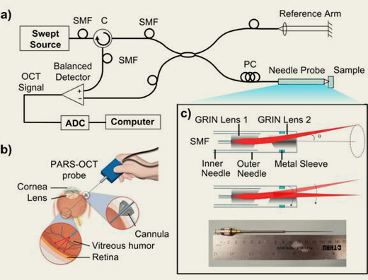
Figure 1. (a) Swept source OCT setup with PARS-OCT probe. (b)
Schematic of the application of PARS-OCT probe during vitrectomy,
a surgical procedure for removal of vitreous humor. (c) Photograph
of PARS-OCT probe and its beam steering principle.
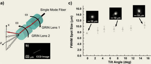
Fig. 2. (a) Planar sweep pattern achieved when
the two GRIN lenses are rotated with equal and opposite velocities.
(b) Scan pattern as recorded by a CCD camera placed 2 mm from
probe tip. (c) Measured spot size (FWHM) vs. tilt angle. Spot
profile is also shown for selected tilt angles.
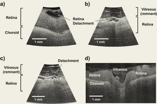
Figure 3. (a, b, and c) Porcine retinal images acquired by
PARS-OCT probe. The retina layer was partially detached during
the removal of vitreous humor. Some remnant vitreouses on the
retina were still visible. (d) Retinal image from a commercial
OCT microscope.
|