Designer
Snowflakes - Part Three
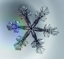
... Precision Snow ... |
|
| The pictures on this page were taken
using the same vapor diffusion chamber as in Part Two,
but with a host of improvements. |
| Better
Snowflakes through Chemistry |
  The first improvement came from my realization that the best
electric needles grow only when certain chemical vapors are added to the diffusion
chamber. Silicone caulk vapor seems to work best, although acetic acid does almost
as well, and even gasoline vapors do pretty well. This trick allowed us to produce
high-quality electric needles much more reliably than before. The first improvement came from my realization that the best
electric needles grow only when certain chemical vapors are added to the diffusion
chamber. Silicone caulk vapor seems to work best, although acetic acid does almost
as well, and even gasoline vapors do pretty well. This trick allowed us to produce
high-quality electric needles much more reliably than before.
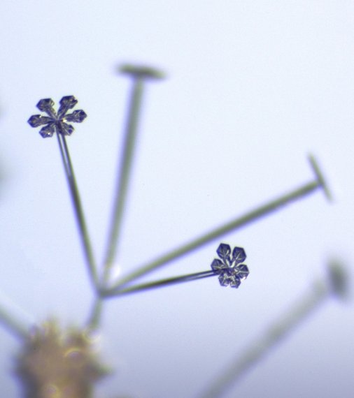
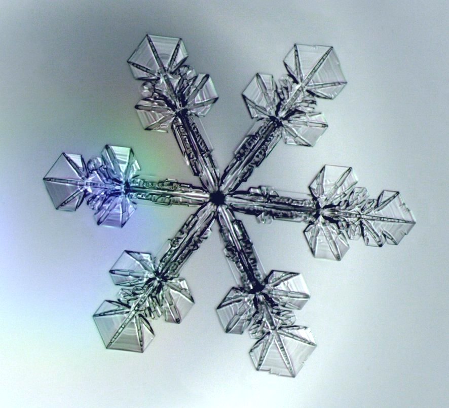
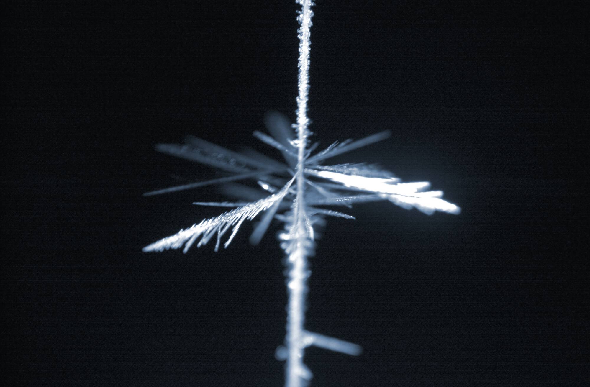
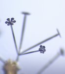
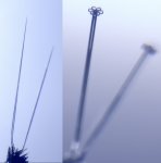
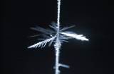
The picture at right shows two near-perfect needles while they were
growing, and after some small stars were grown on their ends. The second images
shows a quartet of electric needles, each with a snow star growing on its end. |
| Snowflakes
on Electric Needles |
  To take advantage of our new
needle-growing technique, we installed a high-power microscope objective right inside the
diffusion chamber, so that we could produce higher-quality pictures of growing snowflakes. To take advantage of our new
needle-growing technique, we installed a high-power microscope objective right inside the
diffusion chamber, so that we could produce higher-quality pictures of growing snowflakes.
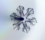
The two images at right are of the same snow crystal at different times; the
left is after 5 minutes of growth, and the right image is after 10 minutes of
growth. The diameter of the larger crystal is about 1.2 mm. The electric
needle is out of focus, behind the crystal on the right side of the image.These were grown with a special manipulator to orient the needles vertically
|
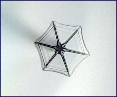
 At left is a simple sectored plate grown at the end of an electric needle;
the plate diameter is about 0.4 mm. To growth such a plate the supersaturation level
needs to be quite low. At left is a simple sectored plate grown at the end of an electric needle;
the plate diameter is about 0.4 mm. To growth such a plate the supersaturation level
needs to be quite low. |
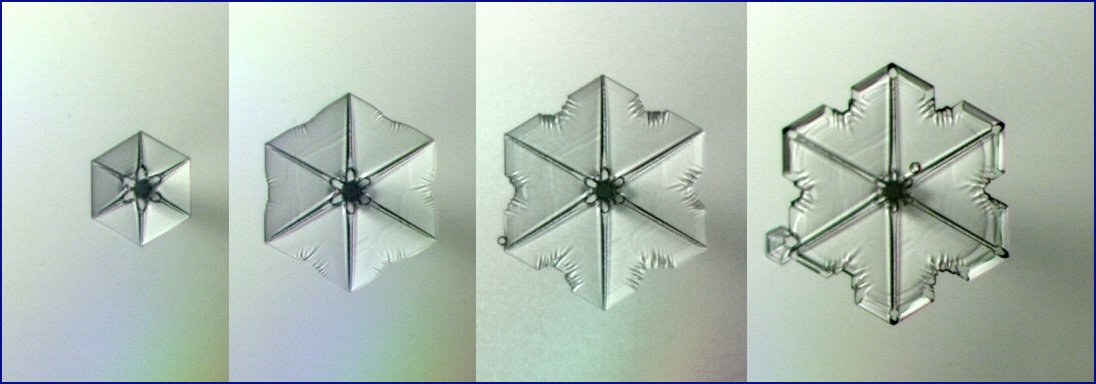
 Another
timeseries showing pictures of a growing plate. The diameter of the final crystal is
about 0.5 mm, and the growth time was about 5 minutes. Between the third and forth
images the temperature was changed, which produced thick edges on the plate. Another
timeseries showing pictures of a growing plate. The diameter of the final crystal is
about 0.5 mm, and the growth time was about 5 minutes. Between the third and forth
images the temperature was changed, which produced thick edges on the plate. |
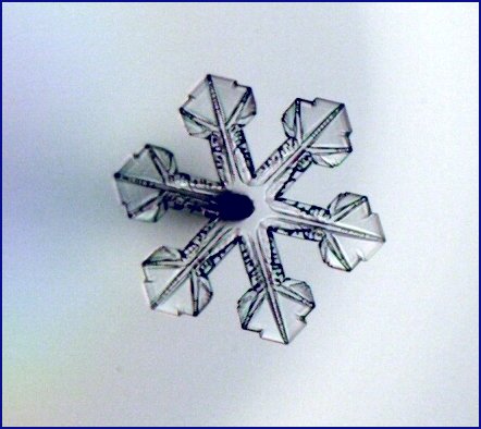
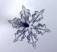  The
two images at right are again pictures of the same crystal at different times; the first
after about 3 minutes of growth, the second after 6 minutes. The diameter of the
larger crystal is about 0.8 mm. The
two images at right are again pictures of the same crystal at different times; the first
after about 3 minutes of growth, the second after 6 minutes. The diameter of the
larger crystal is about 0.8 mm.
|
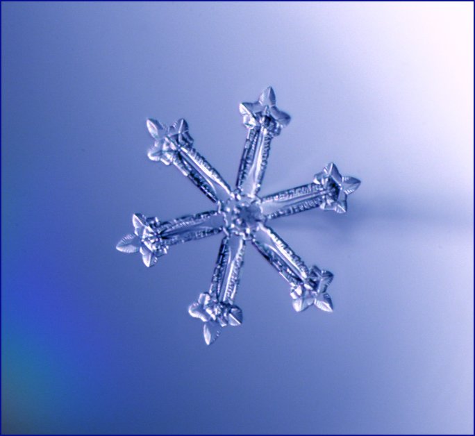
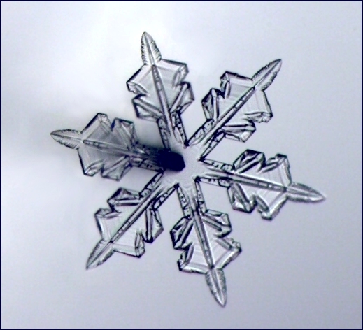
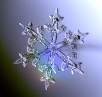
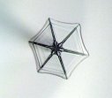
 A
large snow star, for which the electric needle is right behind the crystal and therefore
not visible. The crystal diameter is about 1.5 mm. A
large snow star, for which the electric needle is right behind the crystal and therefore
not visible. The crystal diameter is about 1.5 mm. |
|
| The
World's Largest Snow Crystal? |
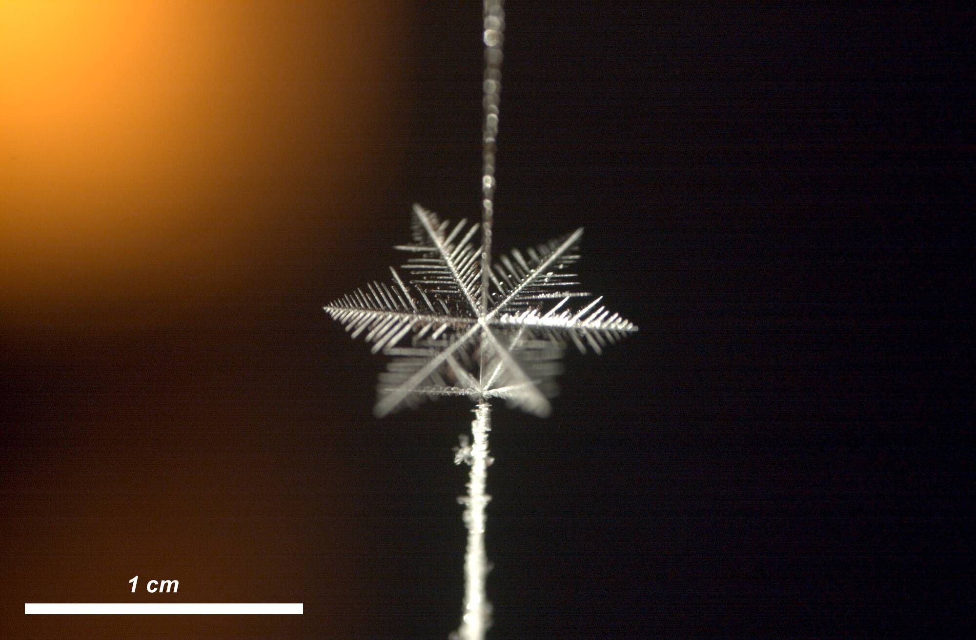
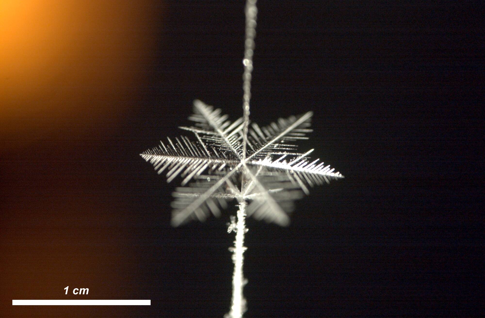
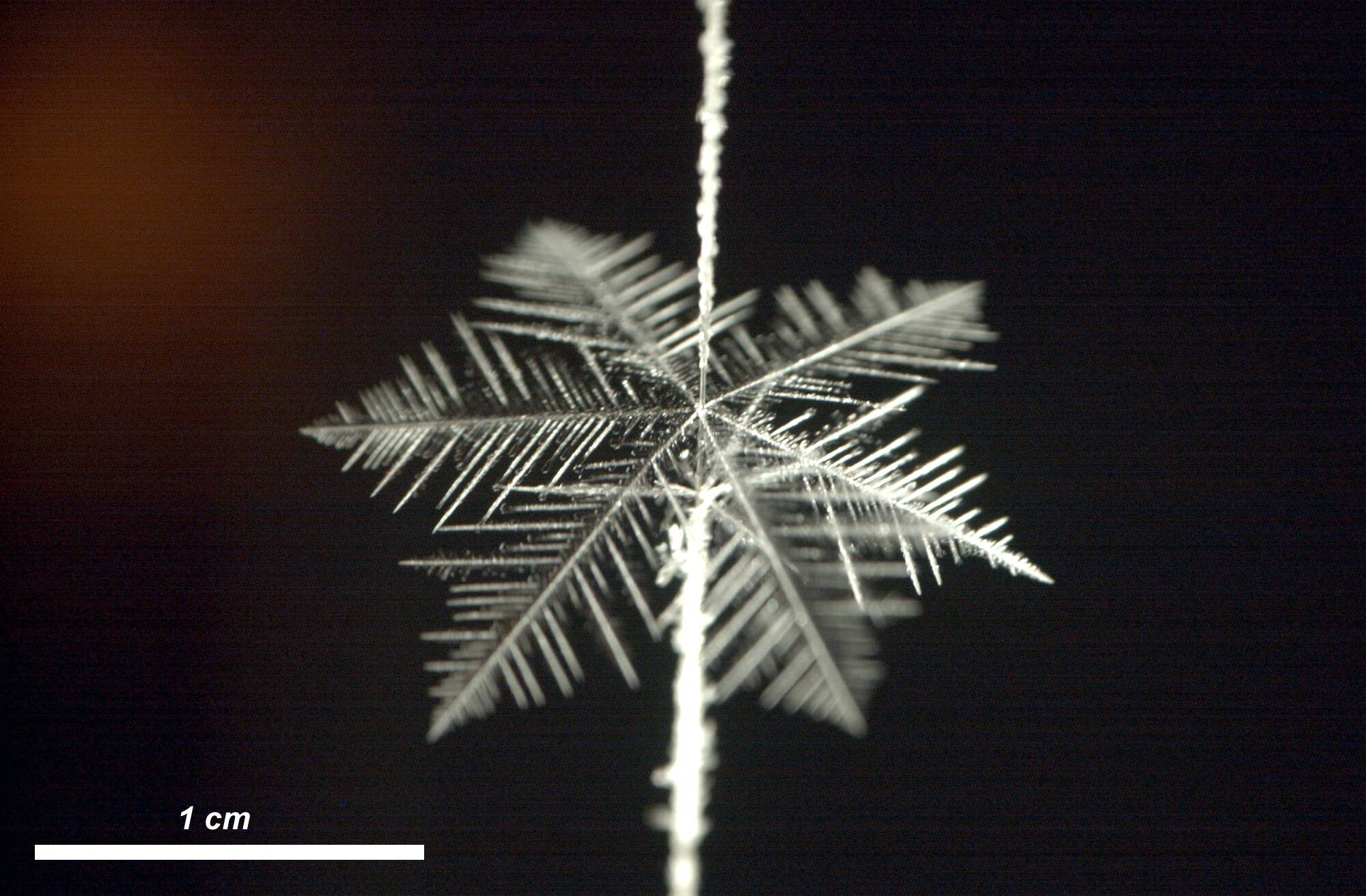
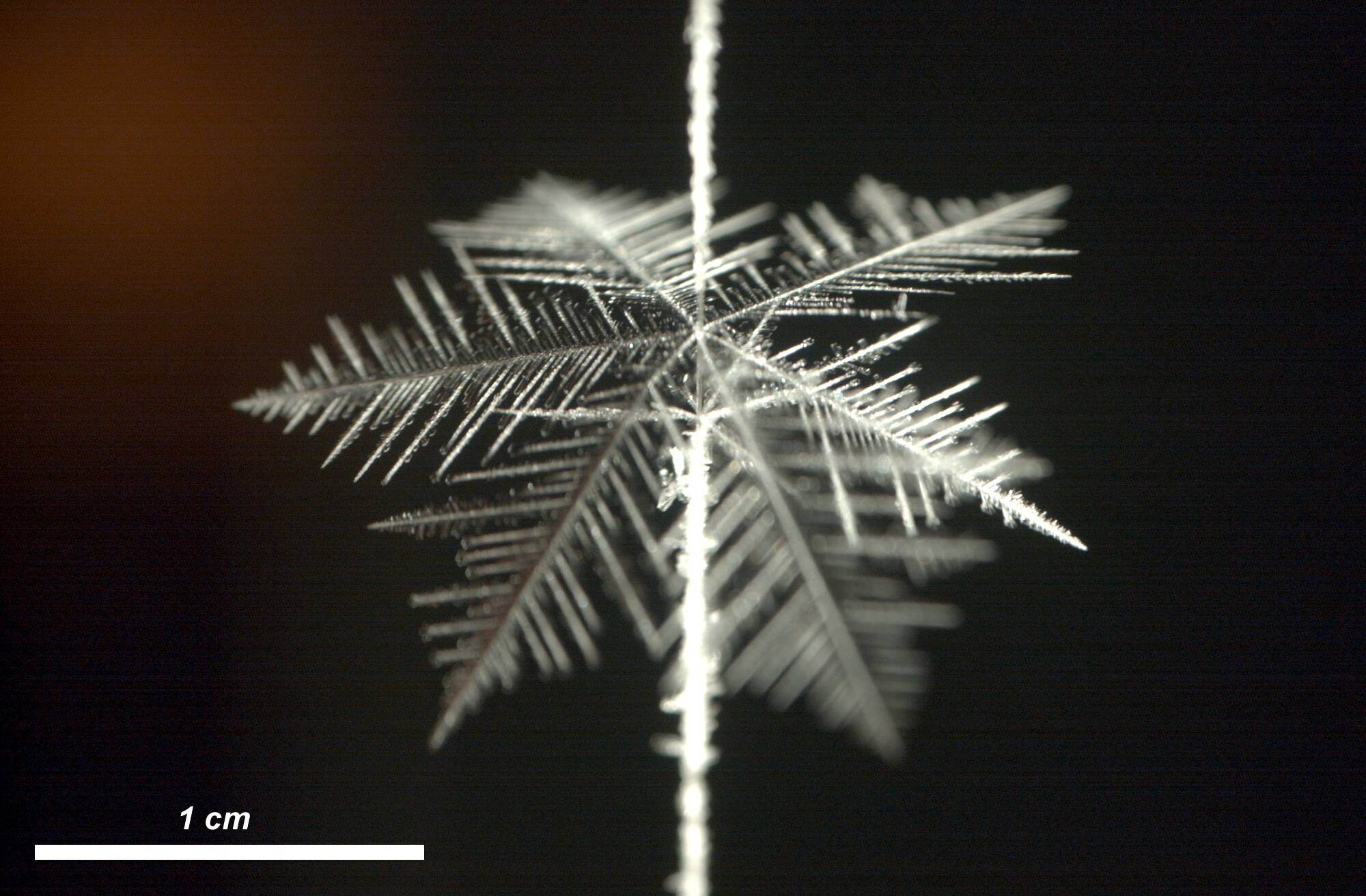
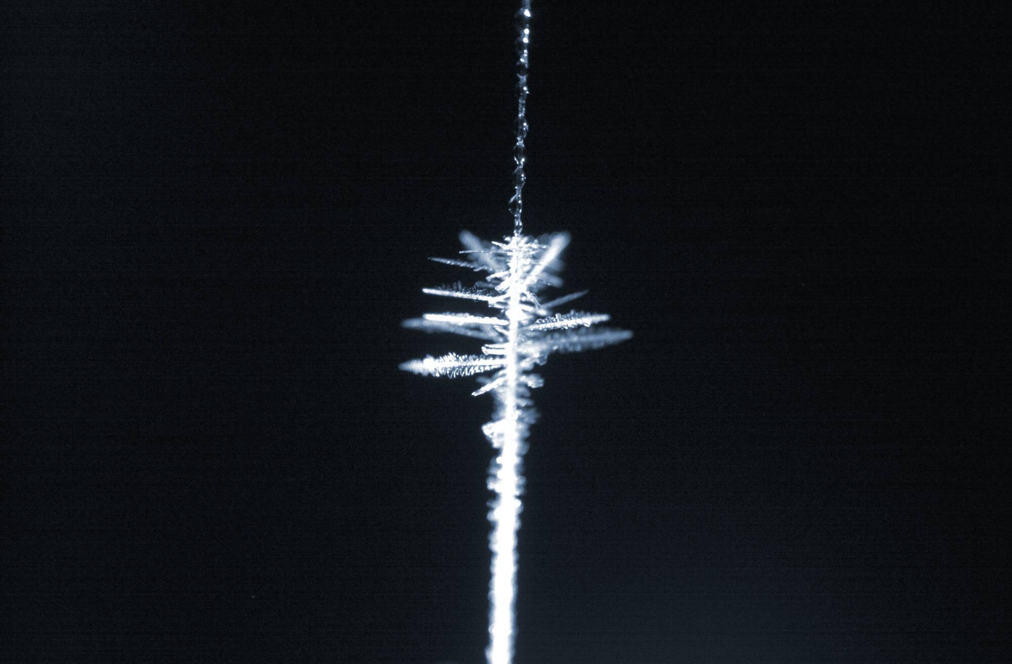
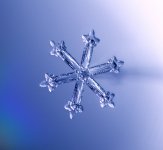
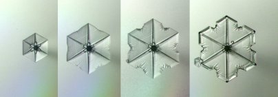
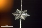
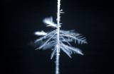
| Below are several
pictures of a very large snow crystal, taken at various times during its growth.
The crystal is clearly a stellar dendrite, which grew on a string in a diffusion chamber
in our laboratory. After about 1.5 hours the crystal had grown to approximately one
inch in diameter, as seen in the final image. |
    |
| |
| Other
Snowflake Structures |
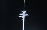
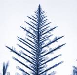
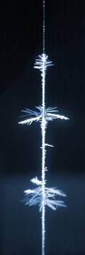 The picture at right is a nice demonstration of
just how strange the growth of ice crystals really is. The image shows crystals
growing on a string hanging in a diffusion chamber. The important feature of a
diffusion chamber is its temperature gradient -- hot at the top and cold at the bottom.
The crystals growing on the string therefore grow at different temperatures.
The top cluster of crystals is growing at -2C, and these crystals grow in the form of
flat, plate-like dendrites. The middle cluster grows at -5C, and these form
needle-like crystals. The bottom cluster grows at -15C, and these too are plate-like
dendrites. The spacing between the top and bottom clusters is 27 mm. Between these
dominant clusters the crystals grow in more blocky shapes, and the growth rates are much
slower. This one picture demonstrates the temperature-dependent crystal growth seen
in the snow crystal morphology diagram (see the Snowflake
Primer). The picture at right is a nice demonstration of
just how strange the growth of ice crystals really is. The image shows crystals
growing on a string hanging in a diffusion chamber. The important feature of a
diffusion chamber is its temperature gradient -- hot at the top and cold at the bottom.
The crystals growing on the string therefore grow at different temperatures.
The top cluster of crystals is growing at -2C, and these crystals grow in the form of
flat, plate-like dendrites. The middle cluster grows at -5C, and these form
needle-like crystals. The bottom cluster grows at -15C, and these too are plate-like
dendrites. The spacing between the top and bottom clusters is 27 mm. Between these
dominant clusters the crystals grow in more blocky shapes, and the growth rates are much
slower. This one picture demonstrates the temperature-dependent crystal growth seen
in the snow crystal morphology diagram (see the Snowflake
Primer).Close-ups of the three different clusters (top,
middle, at lower) are shown in the three images below. One can also see water
droplets above the top cluster, which mark the 0C temperature point on the string. |
   |
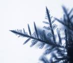
 Even more details of these dominant growth forms are
shown in this and the following three images. The first picture, at left, shows a
crystal grown at a temperature of -15C and high supersaturation (approximately 50
percent). This crystal is plate-like, with well-defined dendritic sidebranches
growing at an angle of 60 degrees from the primary direction. The tip of the
dendrite advanced at a velocity of 2.7 microns/second, along the a-axis of the
crystal. (For this image, and each of the following three images, the enlarged
version is at a scale of two microns/pixel.) Even more details of these dominant growth forms are
shown in this and the following three images. The first picture, at left, shows a
crystal grown at a temperature of -15C and high supersaturation (approximately 50
percent). This crystal is plate-like, with well-defined dendritic sidebranches
growing at an angle of 60 degrees from the primary direction. The tip of the
dendrite advanced at a velocity of 2.7 microns/second, along the a-axis of the
crystal. (For this image, and each of the following three images, the enlarged
version is at a scale of two microns/pixel.)
|
 The next image shows another dendrite, grown in our diffusion chamber at -2C.
The form is quite similar to the -15C dendrite -- a plate-like crystal with
dendritic sidebranching. The tip velocity was 1.2 microns/second, again along the
a-axis. The next image shows another dendrite, grown in our diffusion chamber at -2C.
The form is quite similar to the -15C dendrite -- a plate-like crystal with
dendritic sidebranching. The tip velocity was 1.2 microns/second, again along the
a-axis. |
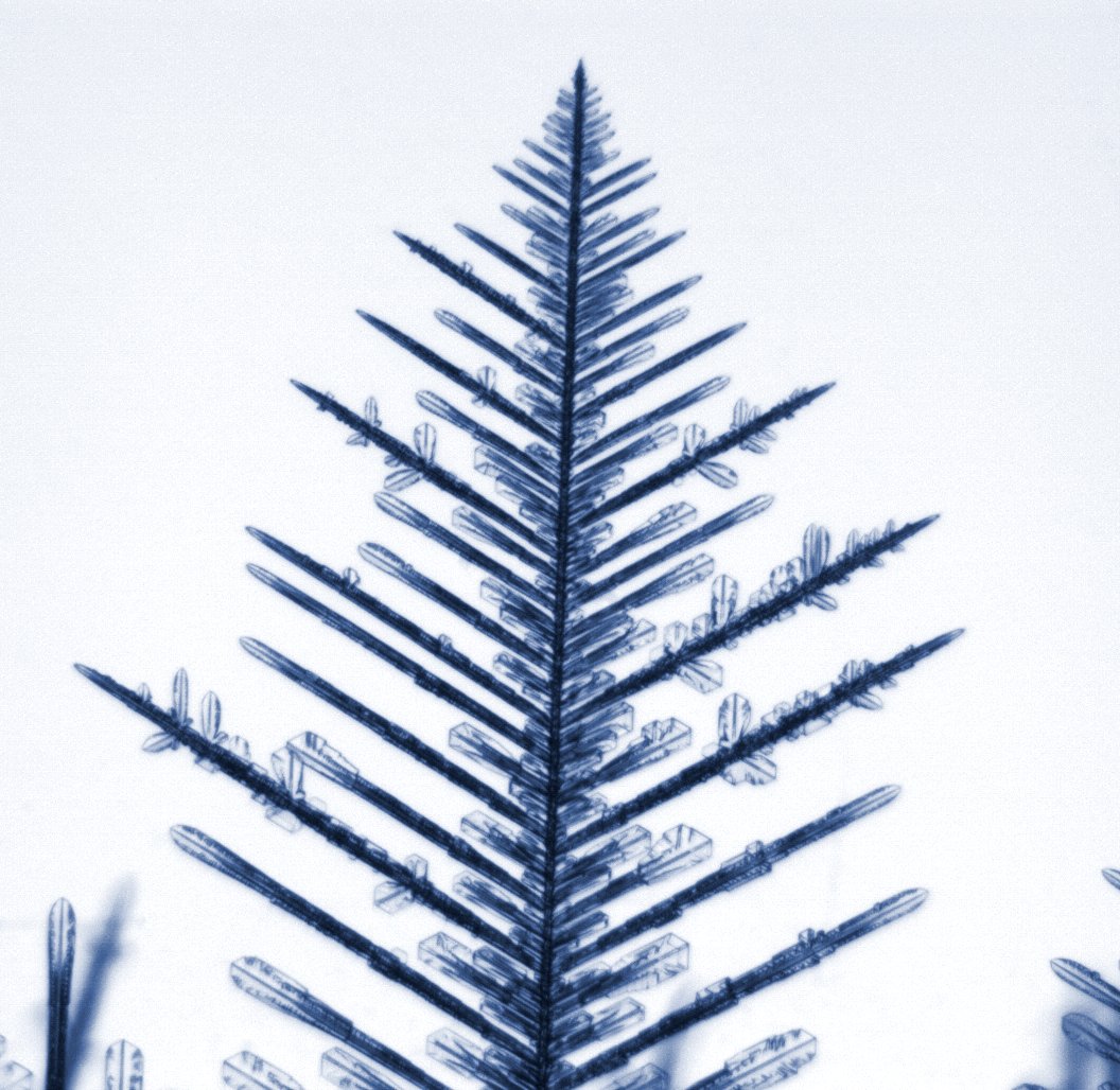
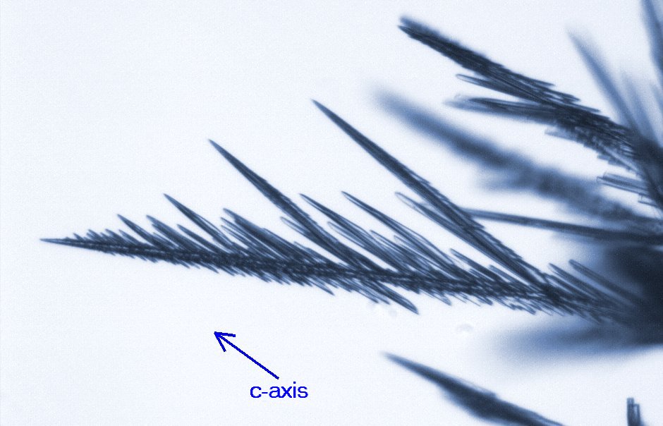
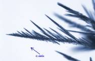  The two images at left are examples of what we call "fishbones" --
needle-like (columnar) crystals that grow at -5C under conditions of high
supersaturation. The left image is the normal growth on a substrate, in this case a
thin wire. The tip of the crystal advanced to the left with a uniform velocity of
2.0 microns/second, but the growth was not along a well-defined crystal axis.
Fishbones exhibit a different kind of sidebranching, with the branches growing roughly
along the c-axis, as shown in the figure. The two images at left are examples of what we call "fishbones" --
needle-like (columnar) crystals that grow at -5C under conditions of high
supersaturation. The left image is the normal growth on a substrate, in this case a
thin wire. The tip of the crystal advanced to the left with a uniform velocity of
2.0 microns/second, but the growth was not along a well-defined crystal axis.
Fishbones exhibit a different kind of sidebranching, with the branches growing roughly
along the c-axis, as shown in the figure.
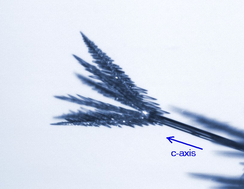
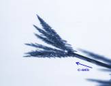
The image on the right shows the start of a set of six fishbones, grown on
the end of an electric needle (see Electric
Growth). The needle grew nicely along the c-axis, thus unambiguously defining
the crystal axes. |