Research
Optofluidic Microscopy
(Primary student researchers: Sean Pang, Seung Ah Lee, Chao Han)
Incorporation of optics into microfluidics has brought forth
a wide spectrum of applications related to imaging, sensing,
spectroscopy, and displays. At present, microfluidics-based imaging
still relies on bulky optical microscopes, although the microfluidic
system itself is usually compact. In Biophotonics lab, we are
developing a compact optical imaging device, termed optofluidic
microscope (OFM). The major component of OFM is a metal coated
CMOS sensor array on which a linear array of subwavelength nanoapertures
is patterned [see Fig. 1].
We can get microscopy level images of an object by stacking it on a high density sensor [see Fig. 2(a)]. The resolution is limited by the sensor pixel size. The OFM system flows the target over a skewed line of apertures that have been etched onto a metal covered sensor array to acquire fine line scans of the object that can then be composited into a high resolution microscope image. In this case, the resolution is equal to the aperture size. Note that the apertures are well separated in this geometry so that they uniquely map onto individual sensor pixels.
Then this device is hermetically sealed
on the floor of a microfluidic delivery channel. The nanoaperture
array is laid down in a slanted fashion under microfluidic channel [see Fig. 2(b)].
We examined our
prototype’s performance by imaging the newly hatched larvae
of C. elegans. A reconstructed image is shown in Fig.
2(d) compared to the direct projection image shown in Fig. 2(e). In addition, high resolution and high throughput of this
initial OFM prototype makes it well suited as a phenotyping device
that can monitor the water quality by imaging the microorganisms, for example, Giardia trophozoites. In Fig 2(f) are some OFM images of of cells and microorganisms.
Also it is possible to construct a phase imaging OFM system by replacing each aperture with 4-hole cluster. By measuring the transmitted interference pattern, we can obtain an amplitude and 2 orthogonal differential phase images of the target [see Fig 2(g)].This new phase imaging technique termed as DIC (differential interference contrast) OFM, has been demostrated in our lab.
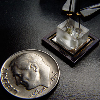
Figure 1. compact OFM prototype, compared with a US dime.
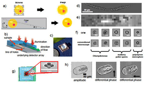
Figure 2. OFM imaging principle b) OFM usage geometry. c) an OFM prototype. d) OFM image of a C. elegans. (1 micron resolution.) e) Image of a C. elegans acquired with an unprocessed CMOS sensor. f) Comparison images of cells and microorganisms. g) A phase imaging OFM system where we have replaced each aperture with 4-hole cluster. h) Starfish larvae images acquired with our offchip demonstration DIC OFM unit.

References
1. Xiquan Cui, Lap Man Lee, Xin Heng, Weiwei Zhong, Paul W. Sternberg, Demetri Psaltis & Changhuei Yang, Lensless high-resolution on-chip optofluidic microscopes for Caenorhabditis elegans and cell imaging, Proceedings of the National Academy of Science Vol 105, 10670 (2008)
2. Xin Heng, David Erickson, Larry R. Baugh, Zahid Yaqoob, Paul W. Sternberg, Demetri Psaltis, and Changhuei Yang. Optofluidic Microscopy: A Method for Implementing High Resolution Optical Microscope On A Chip, Lab on a Chip, DOI:10.1039/b604676b (2006).
3. Jigang Wu, Xiquan Cui, Lap Man Lee & Changhuei Yang, The application of Fresnel zone plate based projection in optofluidic microscopy, Optics Express, Vol. 16 Issue 20, pp.15595-15602 (2008)
4. Xiquan Cui, Matthew Lew & Changhuei Yang, Quantitative differential interference contrast microscopy based on structured-aperture interference, Appl. Phys. Lett. 93 , 091113 (2008)
|