Research
Differential interference contrast (DIC) microscopy based on Youngs interference
Differential interference contrast (DIC) microscopy has been
very successful in rendering excellent phase contrast for transparent
specimens, and is widely used in biology and clinical laboratories.
We are developing a novel DIC microscope based on Young’s
interference, mimicking Young’s original two-slit interference
setup. It does not require any complicated or expensive optical
components, and as such it is less expensive and more robust
than the conventional DIC microscope. In addition, commercially
available DIC microscopes have limitations in imaging the samples
that have anisotropic refractive index distribution such as bones
and teeth. DIC microscopes interfere orthogonally polarized beams
to produce phase contrast. As a result, any change in the linear
polarization caused by the optical anisotropy of the sample can
render an incorrect phase relationship. However our DIC setup
will not suffer from this because our reference and sample beams
have the same polarization. Another promising feature of the
technique is that its simplicity implies that we can easily integrate
it on a chip to implement an on-chip DIC microscope.
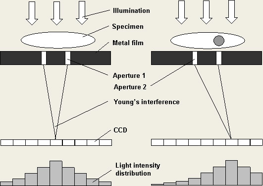
Figure 1. The principle of Differential
interference contrast (DIC) microscopy based on Young’s
interference
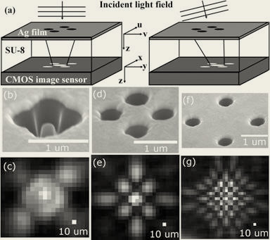
Figure 2. (a) Device
geometry and principle of operation. (b) SEM image and (c)
interference pattern of 600 nm holes with 600 nm spacing. (d),
(e) Same plots for holes with 1.2 μm spacing. (f), (g) Same plots for holes
with 2.4 μm spacing.
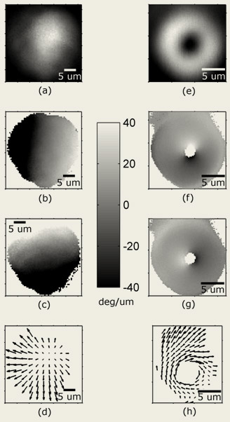
Figure 3. (a) Intensity, (b) u component
and (c) v component of differential phase, and (d) vector representation
of differential phase of a Gaussian beam. (e)–(h) Same
plots for an optical vortex.

References
M. Lew, X. Q. Cui, X. Heng, and C. H. Yang, "Interference
of a four-hole aperture for on-chip quantitative two-dimensional
differential phase imaging," Optics Letters 32, 2963-2965
(2007). (pdf)
|