When it comes to quantum technologies, computing has dominated headlines around the world. Computers that exploit the laws of quantum mechanics are significantly faster for several classes of problem than even the most powerful supercomputers..." More>>
Researchers in Caltech's Andrew and Peggy Cherng Department of Medical Engineering have made a major step forward in medical imaging by taking inspiration from the field of astronomy..." More>>
Photoacoustic microscopy (PAM) is a relatively new imaging technique that uses laser light to induce ultrasonic vibrations in tissue. These ultrasonic vibrations, along with a computer that processes them, can then be used to create an image of the structures of the tissue in much the same way ultrasound imaging works...." More>>
Reach out right now and touch anything around you. Whether it was a key on your keyboard, the wood of your desk, or the fur of your dog, you felt it the instant your finger contacted it..." More>>
Of the many ways to treat cancer, the oldest, and maybe most tried and true, is surgery. Even with the advent of chemotherapy, radiation therapy, and more experimental treatments like bacteria that seek and destroy cancer cells, cancers, very often, simply need to be cut out of a patient's body..." More>>
In an effort to create more opportunities for students, increase interdisciplinary research, and gain visibility for a first-of-its kind program, Caltech is creating a new graduate education track that combines medical engineering and electrical engineering..." More>>
The field of PA imaging for human clinical use reached a substantial milestone in 2021, with FDA approval for a platform using PA and ultrasound technologies in tandem to differentiate between benign and malignant breast lesions..." More>>
When something happens quickly, we say it takes place in the blink of an eye. Yet, during the 0.1 seconds it takes to blink, a camera developed by Lihong Wang can take 7 trillion pictures..." More>>
Keck School of Medicine of USC researchers, working with a Caltech team, demonstrate possibilities of innovative imaging technology to visualize brain function..." More>>
A Caltech professor, in collaboration with researchers at the University of Southern California, has demonstrated for the first time a new technology for imaging the human brain using laser light and ultrasonic sound waves...." More>>
There are things in life that can be predicted reasonably well. The tides rise and fall. The moon waxes and wanes. A billiard ball bounces around a table according to orderly geometry..." More>>
Two Caltech faculty members, Lihong Wang and Changhuei Yang, have been named fellows of the National Academy of Inventors (NAI). According to the NAI, election as a fellow is the "highest professional distinction accorded to academic inventors who have demonstrated a prolific spirit of innovation in creating or facilitating outstanding inventions that have made a tangible impact on quality of life, economic development and the welfare of society..." More>>
Two Caltech researchers have received funding for neuroscience projects from the National Institutes of Health's Brain Research through Advancing Innovative Neurotechnologies (BRAIN) Initiative... More>>
A new camera that takes videos at record-breaking speeds of up to 100 billion frames per second in 3D has been demonstrated by researchers at the California Institute of Technology in the US. The feat was made possible by a technique known as single-shot stereo-polarimetric compressed ultrafast photography (SP-CUP), and it builds on the groups earlier work including a camera that takes images at 70 trillion frames per second, which is fast enough to see light travel... More>>
In January this year, Lihong Wang combined his worlds fastest camera with phase contrast microscopy to image ultrafast phenomena in transparent objects at a blisteringly fast one trillion frames per seconds... More>>
In his quest to bring ever-faster cameras to the world, Caltech's Lihong Wang has developed technology that can reach blistering speeds of 70 trillion frames per second, fast enough to see light travel. Just like the camera in your cell phone, though, it can only produce flat images... More>>
For as much as cameras allow us to experience phenomena that would otherwise go unnoticed, their imaging speeds still fundamentally limit our capability to see, well, everything. Now, scientists at the California Institute of Technology hope to change that... More>>
Just about everyone has had the experience of blinking while having their picture taken. The camera clicks, your eyes shut, and by the time they open again, the photo is ruined. A new ultrafast camera developed at Caltech, were it aimed at your lovely face, could also capture you looking like a dunce with your eyes shut, except instead of taking just one picture in the time it takes you to blink, it could take trillions of pictures... More>>
The best phone cameras can record slow motion with under 1,000 frames per second. Commercial rigs generally shoot with several thousand. But that all absolutely pales in comparison to the new record holder for the worlds fastest camera, boasting a mind-boggling rate of 70 trillion frames per second. Thats fast enough to capture light waves in movement... More>>
Photoacoustic imaging, a technique for examining living materials through the use of laser light and ultrasonic sound waves, has many potential applications in medicine because of its ability to show everything from organs to blood vessels to tumors... More>>
US-based researchers have combined extreme speed photography with phase contrast microscopy to image ultrafast phenomena in transparent objects, from cells to shockwaves, at picosecond resolution... More>>
New camera technology that takes up to 1 trillion frames per second is so advanced it can take images of transparent phenomena, U.S. researchers say. The camera builds on previous research, in which the team used the technology to capture light traveling in slow motion... More>>
A little over a year ago, Caltech's Lihong Wang developed the world's fastest camera, a device capable of taking 10 trillion pictures per second. It is so fast that it can even capture light traveling in slow motion... More>>
Label-free mid-infrared (MIR) microscopy provides rich chemical and structural information on biological tissues. While traditional histopathology requires time-consuming tissue section processing and staining, MIR microscopy can obtain histopathologic information without staining. However, strong MIR absorption of water in fresh biological samples results in high background and low contrast, which limits conventional MIR techniques to imaging only dried and thin-sliced tissue specimens. Moreover, at long MIR wavelengths, optical diffraction severely limits lateral resolution, preventing the technique from resolving subcellular information. This year, we developed ultraviolet-localized MIR photoacoustic microscopy (ULMPAM), which can achieve high-resolution and water backgroundfree MIR imaging of fresh biological samples... More>>
If you were to use your smartphone to record a video you are talking about maybe a 30 Hertz frame rate, meaning you capture 30 frames per second. That is good enough for daily phenome-non but if you want to capture something much faster, we have to do something very different. Of course the fastest phenomenon in the world is light propagation. Light pulses propagate at the speed of light and as we know that's the terminal speed, nothing can propagate faster than the speed of light. The fastest 1D camera is some-thing called streak camera that gives you 1 T images but at a very high, not frame but, line rate, so we want to add one more dimension to it... More>>
Targeting medical treatment to an ailing body part is a practice as old as medicine itself. A Band-Aid is placed on a skinned knee. Drops go into itchy eyes. A broken arm goes into a cast. But often what ails us is inside the body and is not so easy to reach. In such cases, a treatment like surgery or chemotherapy might be called for. A pair of researchers in Caltech's Division of Engineering and Applied Science are working on an entirely new form of treatmentmicrorobots that can deliver drugs to specific spots inside the body while being monitored and controlled from outside the body... More>>
It may look like a giant game of space invaders, but each colored dot in this image is an individual cancer cell within a droplet of blood. In this scanned image of single-cell metabolic photoacoustic microscopy (SCM-PAM), different colors represent the amount of oxygen dissolved in each well. To measure oxygen consumption rate, researchers place cells into individual wells filled with blood; those with higher metabolisms consume more oxygen, lowering the level remaining in the blood... More>>
Researchers in Lihong Wang's lab have developed a new imaging technique that uses pulses from two kinds of lasers to take pictures of microscopic biological structures... More>>
The oxygen consumption rates of single tumour cells can be measured via photoacoustic microscopy, by leveraging haemoglobin as both an oxygen supplier and an oxygen sensor... More>>
Photoacoustic computed tomography (PACT) is a non-invasive hybrid imaging technique that excites biological tissues with light and detects the subsequently generated ultrasound to form images. PACT combines the advantages of both optical imaging--high optical contrast, and ultrasonic imaging--high resolution and deep penetration in biological tissues. PACT has been widely used for vascular network mapping, functional brain imaging, and tumor detection in deep tissues... More>>
Devising the best treatment for a patient with cancer requires doctors to know something about the traits of the cancer from which the patient is suffering. But one of the greatest difficulties in treating cancer is that cancer cells are not all the same. Even within the same tumor, cancer cells can differ in their genetics, behavior, and susceptibility to chemotherapy drugs... More>>
Tumours consist of heterogeneous populations of cancer cells that have distinct genetic and phenotypic profiles. Cellular heterogeneity within a tumour, namely intratumoural heterogeneity, has become a great barrier to effective cancer therapy. Assessing the extent of intratumoural metabolic heterogeneity would greatly contribute to our understanding of tumour growth, invasion, and drug resistance... More>>
In 2003, Lihong Wang, then at Texas A&M University, US, delivered images of brains in living rats that well and truly moved the field of photoacoustic imaging into the fast lane. Revealing intricate networks of blood vessels in the rat cerebral cortex - acquired with the skin and skull intact - the tomograms represented a massive leap forward for functional imaging using photoacoustics, also known as optoacoustics, and stunned researchers worldwide... More>>
Rooted in the physics of space-time duality, temporal focusinga time-domain counterpart of spatial focusinghas found diverse applications in nonlinear microscopy and materials processing. Temporal-focusing events are often non-repeatable, which precludes scrutinizing the propagation of ultrashort laser pulses in living biological tissue and investigating the elusive physics of strong-field interactions with matter... More>>
New scanners on the market are providing higher image quality with faster acquisition time, driving demand among cardiology and oncology departments. The systems are being packaged with new and improved applications to simplify the exam process and radiation dose optimization technology to ensure patients are being imaged in the safest and most efficient ways possible... More>>
Single-shot 10-trillion-frame-per-second compressed ultrafast photography (CUP) is now possible with a new camera, developed by researchers from Institut National de la Recherche Scientifique (INRS) and California Institute of Technology (Caltech). The camera system, called T-CUP, passively captures dynamic events with 100-femtosecond (fs) frame intervals in a single camera exposure. According to the researchers, T-CUP has set a new record for real-time imaging speed, even capturing light in extremely slow motion... More>>
Additional media coverage of our recent 10-trillion-frame-per-second CUP Camera can be found here>>
In recent years, the junction between innovations in non-linear optics and imaging has opened the door for new and highly efficient methods for microscopic analysis of dynamic phenomena in biology and physics. But to harness the potential of these methods, there needs to be a way to record images in real time at a very short temporal resolutionin a single exposure... More>>
What happens when a new technology is so precise that it operates on a scale beyond our characterization capabilities? Although some measurements are possible, nothing beats a clear image, says INRS professor and ultrafast imaging specialist Jinyang Liang. He and his colleagues, led by Caltech's Lihong Wang, have developed what they call T-CUP: the world's fastest camera, capable of capturing 10 trillion frames per second. This new camera literally makes it possible to freeze time to see phenomenaand even lightin extremely slow motion... More>>
Our goal is to build a dream machine for breast screening, diagnosis, monitoring, and prognosis without any harm to the patient. We want it to be fast, painless, safe, and inexpensive... More>>
Researchers have been developing a new method for detecting breast cancer called photoacoustic computed tomography (PACT). The technique uses harmless pulses of laser light to penetrate the breast tissue. This causes a type of sound wave called photoacoustic waves to spread through the tissue. These waves can be measured by sensors surrounding the breast... More>>
The image shows a scan of a human breast performed by photoacoustic computed tomography (or PACT) a new imaging technology that may one day replace the typical X-ray mammograms that women over the age of 40 routinely undergo to check for the presence of breast cancer... More>>
Knowing that the conventional method for breast cancer screeningmammographyis less than ideal for multiple reasons (including exposure to x-ray radiation and discomfort for patients), researchers at the California Institute of Technology (Caltech; Pasadena, CA) have developed a laser-sonic scanner that can find tumors in as little as 15 seconds by shining pulses of light into the breast... More>>
Early detection has been shown to increase breast cancer survival rates, but many women avoid having their mammograms taken as often as they should because of the discomfort involved. A 2013 study found that as many as half of women who were avoiding their mammograms cited pain as the reason why... More>>
Researchers from the California Institute of Technology (Caltech) in Pasadena have developed a single-breath-hold photoacoustic CT (SBH-PACT) system that can image a patients breast in 15 seconds and requires no ionizing radiation or contrast agents, sharing their findings in a new study published by Nature Communications... More>>
Up to 50% of women skip potentially life-saving mammograms often because the procedure can cause extreme discomfort and pain. Now researchers have developed a painless, light-based, non-radioactive, 15-second procedure that could revolutionize breast cancer screening and save lives... More>>
Caltech researchers say they have developed something better: a laser-sonic scanner that can find tumors in as little as 15 seconds by shining pulses of light into the breast... More>>
The Optical Society (OSA) is pleased to name Lihong Wang, California Institute of Technology, USA, the 2018 Michael S. Feld Biophotonics Award recipient. Wang is recognized for inventing the worlds fastest two-dimensional receive-only camera and enabling real-time imaging of the fastest phenomena such as light propagation and fluorescence decay... More>>
In the first anniversary of the launch of Nature Biomedical Engineering, the editors have selected 10 pieces of content that exemplify the interdisciplinarity of the subject area and the need for closer collaboration between bench researchers, clinicians and medical engineers to solve outstanding health challenges... More>>
Lihong Wang, Bren Professor of Medical Engineering and Electrical Engineering, has been elected to the National Academy of Engineering (NAE). Election to the NAE is considered among the highest professional distinctions in the engineering field... More>>
Lihong V. Wang, who earned his Ph.D. in electrical engineering while working with two Nobel Laureates at Rice University, has been elected to the National Academy of Engineering (NEA)... More>>
Caltech engineers have improved a technique for taking three-dimensional (3-D) microscopic images of tissue, allowing them to see inside living creatures with greater precision than before... More>>
In this video, Barbara Gefvert, editor-in-chief of BioOptics World, speaks with Lihong Wang, Ph.D., Bren Professor of Medical Engineering and Electrical Engineering at Caltech, about his exciting work in photoacoustic imaging for peering noninvasively into biological tissues... More>>
Small animals such as rodents are indispensable models for preclinical and basic science research. Seeing inside their entire bodies with high spatial and time resolution could provide insights into biological processes, advancing studies of human disease and drug development... More>>
Photoacoustic computed tomography (PACT) is an optical imaging technology used for whole-body imaging of small laboratory animals. A new single-impulse panoramic PACT (SIP-PACT) system is enabling researchers to perform high-resolution in vivo imaging of whole-body dynamics of small animals in real time. The system combines high spatiotemporal resolution, deep penetration, anatomical, dynamical and functional contrasts, and full-view fidelity... More>>
For many patients diagnosed with cancer, the primary treatment plan is surgery to remove the tumor, but in the quest to retain as much healthy tissue as possible, it's impossible for surgeons to know if they've removed all traces of the cancerous tissue while in the operating room. A new imaging technique would give doctors this ability, thus avoiding having the patient return to surgery weeks later for a second procedure... More>>
Engineers at the Optical Imaging Laboratory led by Caltech's Lihong Wang have developed an imaging technology that could help surgeons removing breast cancer lumps confirm that they have cut out the entire tumor--reducing the need for additional surgeries... More>>
Medical engineers at the Optical Imaging Laboratory led by Caltech's Lihong Wang are now able to take a live look at the inner workings of a small animal with enough resolution to see active organs, flowing blood, circulating melanoma cells, and firing neural networks... More>>
Using a combination of light and sound, Lihong Wang is noninvasively peering deeper inside biological tissues than previously possible. Three-dimensional photoacoustic microscopy and functional photoacoustic computed tomography, the technologies first reported by WangCaltech's Bren Professor of Medical Engineering and Electrical Engineeringgenerate detailed color images of tumors and other structures inside the body... More>>
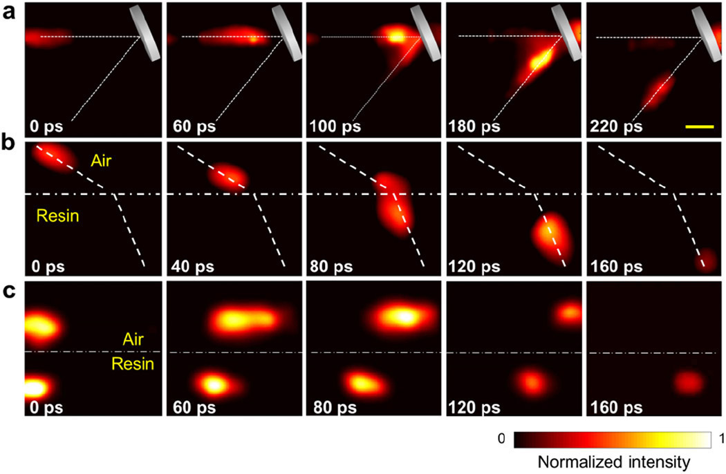
In this video interview, Lihong Wang of Calltech discusses a single-shot ultrafast video recording of a light-induced photonic Mach cone propagating in an engineered scattering plate assembly. This dynamic light-scattering event was captured in a single camera exposure by lossless-encoding compressed ultrafast photography at 100 billion frames per second...More>>
Ultrafast two-dimensional imaging of dynamic phenomena in real time should ideally achieve a picosecond-level exposure time per frame while avoiding temporal and spatial scanning. Now, Jinyang Liang and collaborators have successfully recorded light-scattering dynamics in the form of a photonic Mach cone with a single camera exposure at a rate of 100 billion frames per second...More>>
This is the fastest race in the Universe, at nearly 300,000 km/s ... and yet it did not stop the physicists of Lihong Wang's team from the University Of Washington, in St Louis (USA), to film for the first time the propagation of light in a vacuum...More>>(French)
Biomedical engineers in the US have developed a form of photoacoustic imaging that can quantify the elasticity of human tissue. The technique, which the engineers tested on skeletal muscle in a human, could be used to monitor the elasticity of the cervix during pregnancy, for example, potentially allowing doctors to predict premature delivery dates...More>>
Researchers have improved upon a new camera technology that can image at speeds about 100 times faster than today’s commercial cameras while also capturing more image frames. The new technology opens a host of new possibilities for studying extremely fast processes such as neurons firing, chemical reactions, fuel burning or chemicals exploding...More>>Digital Journal, Laser Focus World,
TMCnet, ITbriefing,
4-Traders
In an ordinary digital camera, a large amount of information is collected by the camera’s sensor. This information is immediately processed by compression software, and a much smaller amount is then stored. This process takes time, contributing largely to what is...More>>
A new approach to high-speed photography could help capture the clearest-ever footage of light pulses, explosions or neurons firing in the brain, according to a team of ultrafast camera developers. The technique involves shooting 100 billion frames per second in a single exposure without an external light source. That means, for example, there would be no need to set off multiple explosions just to gather enough data to create a video reconstructing exactly how chemicals react to create the blast...More>>
PBS NewsHour: This camera snaps photos 3 billion times faster
than an iPhone. A new approach to high-speed photography could
help capture the clearest-ever footage of light pulses, explosions or
neurons firing in the brain, according to a team of ultrafast camera
developers. The technique involves shooting 100 billion frames per
second in a single exposure without an external light source. That
means, for example, there would be no need to set off multiple
explosions just to gather enough data to create a video reconstructing
exactly how chemicals react to create the blast...More>>
Dr. Lihong Wang will give a talk in the upcoming 2016 Gordon Research Conference on Image Science. The time and the placeholder title for his presentation is:
Lihong Wang
Session: Imaging at the Physical Limits
Time: Tuesday morning, June 7
Placeholder title: Redefining the spatiotemporal limits of optical imaging
Direct visualization of transient events holds great interest in physics, chemistry and biology. Existing CCD or CMOS detectors have limited on-chip storage and data readout speeds, however, and so cannot image events lasting less than 1 ns. The streak camera—an ultrafast imager that converts time to space by pulling photoelectrons with a sweeping voltage along the axis perpendicular to the device’s entrance slit—breaks that speed limit. Yet the streak camera conventionally is a 1-D imager, acquiring one line of the scene per measurement. That makes mechanical scanning necessary to obtain a 2-D image, so the transient event must be reproducible for repetitive measurements...More>>
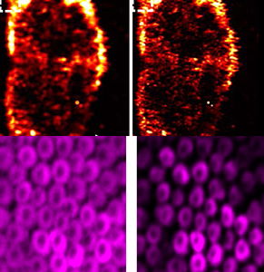
The researchers – from Lihong Wang's laboratory at Washington University in St. Louis and Vladislav Verkhusha's laboratory at the Albert Einstein College of Medicine – genetically modified U87 human glioblastoma cells to express BphP1, a bacterial phytochrome protein cloned from a purple photosynthetic bacterium Rhodopseudomonas palustris. BphP1 exhibits natural photochromic behaviour, switching between two absorbing states: from an "ON" state to an "OFF" state upon 730–790 nm illumination, and back upon 630–690 nm illumination...More>>
A team of engineers, led by Lihong Wang, PhD, and postdoctoral researcher Junjie Yao, found a way to clearly see tiny amounts of live cancer cells as deep as 1 centimeter in tissue using photoacoustic tomography. They did so by genetically modifying glioblastoma cancer cells to express BphP1 protein, derived from a bacterium commonly found in soil and water...More>>
Researchers continue to search for solutions to the problem of focusing light in scattering media—an essential requirement for optical imaging of biological tissues at any meaningful depth. A team of scientists from Washington University of St. Louis (USA) now reports that it has devised an improvement to wavefront-shaping techniques that can substantially boost the speed, quality, and practicality of imaging in scattering media. The team sees applications for the technique ranging from deep-tissue biophotonic imaging to real-time capture of movement and flow of blood cells in living tissue...More>>
Wang, a professor of biomedical engineering at Washington University in St. Louis, has already helped develop instruments that can detect individual cancer cells in the bloodstream and oxygen consumption deep within the body. He has also created a camera that shoots at 100 billion frames a second, fast enough to freeze an object traveling at the speed of light...More>>
To minimize complexity, researchers often study cellular proteins or nucleic acids in isolation. But sometimes—when testing a drug’s efficacy and safety, for instance, or monitoring tumor progression—ex vivo just won't do. The only way to know how a compound or cells will behave in the body is to put them into an animal and watch what happens live. The results are easily recognizable in the pages of your favorite journal: the ghostly outline of a mouse, with a telltale multicolored heat bloom indicating where the action is...More>>
A human skull, on average, is about 0.3 inches thick, or roughly the depth of the latest smartphone. Human skin, on the other hand, is about 0.1 inches, or about three grains of salt, deep.
While these dimensions are extremely thin, they still present major hurdles for any kind of imaging with laser light.
Why? Laser light contains photons, or miniscule particles of light. When photons encounter biological tissue, they scatter. Corralling the tiny beacons to obtain meaningful details about the tissue has proven one of the most challenging problems laser researchers have faced.
However, one research group at Washington University in St. Louis (WUSTL) decided to eliminate the photon roundup completely and use scattering to their advantage...More>>
A team of biomedical engineers at Washington University, led by Dr. Lihong Wang, has developed the world’s fastest receive-only 2-D camera. The device can capture events up to 100 billion frames per second. The camera tracks light, capturing images of a single laser pulse, opening the door for scientific exploration and new discoveries...More>>
Newswise — An NIBIB grantee has developed an ultrafast camera that can acquire two-dimensional images at 100 billion frames per second, a speed capable of revealing light pulses and other phenomena previously too fast to be observed.
“When you turn on a laser pointer, you see an immediate beam of light. That’s because light moves so fast, you aren’t able to detect its movement with the naked eye. Using this camera, light is revealed as traveling through space from one point to another,” says the camera’s inventor, Lihong Wang, Ph.D., a professor of biomedical engineering at Washington University in St. Louis...More>>
Video recording of ultrafast phenomena such as dynamic events in molecular biology would transform our understanding of a range of phenomena. However, using a detector array based on CCD or CMOS technologies is fundamentally limited by the sensor's on-chip storage and data transfer speed. To get around this problem, the most practical approach is to use a streak camera. In this ultrafast imaging device, the incident light first passes through a narrow entrance slit (usually 50µm wide) and is imaged onto the photocathode of a streak tube. Here, the incident light is converted to photoelectrons, which are accelerated by an accelerating mesh. A pair of electrodes then applies a sweeping (i.e., time-varying) voltage along the axis perpendicular to the device's entrance slit. Because of this sweeping voltage, electrons arriving at different times are deflected to different spatial positions, and these electrons are then multiplied by a microchannel plate. They subsequently bombard a phosphor screen and are converted back into light. The phosphor screen is imaged to a CCD, which records the image. However, the resultant image is normally 1D: only a single line of the scene can be seen at a time. Acquiring a 2D image requires mechanical scanning across the entire field of view, which poses severe restrictions on the recordable scenes because the event itself must be repetitive...More>>
Researchers studying cancer and other invasive diseases rely on high-resolution imaging to see tumors and other activity deep within the body’s tissues. Using a new high-speed, high-resolution imaging method, Lihong Wang, PhD, and his team at Washington University in St. Louis were able to see blood flow, blood oxygenation, oxygen metabolism and other functions inside a living mouse brain at faster rates than ever before.
Using photoacoustic microscopy (PAM), a single-wavelength, pulse-width-based technique developed in his lab, Wang, the Gene K. Beare Professor of Biomedical Engineering in the School of Engineering & Applied Science, was able to take images of blood oxygenation 50 times faster than their previous results using fast-scanning PAM; 100 times faster than their acoustic-resolution system; and more than 500 times faster than phosphorescence-lifetime-based two-photon microscopy (TPM)...More>>
Shielded by the skull and packed with fatty tissue, the living brain is perhaps the most difficult organ for scientists to probe. Functional magnetic resonance imaging (fMRI), which noninvasively measures changes in blood flow and oxygen consumption as a proxy for neuronal activation, lags far behind the actual speed of thought. Now, a new technique may provide the fastest yet method of measuring blood flow in the brain, scientists report online today in Nature Methods. The technique, which bounces laser beams off red blood cells, has a resolution of under a millisecond—slightly less time than it takes a neuron to fire—and it has a far higher spatial resolution than fMRI. Even the most powerful fMRI machines, used only on animals, can image only millimeter-wide swaths of tissues including thousands of cells. The new technique, which takes its measurements from sonic waves produced by the beams, can image structures as small as individual blood vessels and cells (see above). Although the technique is not likely to be feasible in humans due to safety concerns, it could provide an important tool to better understand how blood flow and oxygen consumption is related to brain activity. That’s a key question for those relying on cruder and safer tools, such as fMRI, to study the human brain, researchers say. It is also a powerful tool for studying how errant eddies and whorls of blood in blood vessels can sometimes lead to stroke, they say.
Exploiting a strategy known as compressed sensing, the camera can capture video at 100 billion frames per second.
The human visual system can perceive only about 10 distinct images per second. So to record everyday life as we experience it, a standard video capture rate of 24 frames per second (fps) more than suffices. But many physical phenomena unfold faster than the eye can see. To record them, researchers seek ever-faster cameras.
Now Lihong Wang and coworkers at Washington University in St. Louis have developed a camera that can record at 1011 fps, good enough to capture the movement of light at millimeter length scales.1 The camera marries streak photography, an ultrafast imaging technique previously limited to filming in one spatial dimension, with compressive sensing, a mathematical strategy for reconstructing an entire scene from an incomplete set of measurements. “The camera has countless potential applications,” comments Mário Figueiredo of Instituto Superior Técnico in Lisbon, Portugal. “It’s sort of like a microscope for time.”...More>>
Using techniques adapted from astronomy, physicists are finding ways to see through opaque materials such as living tissue...
“Just ten years ago, we couldn't imagine high-resolution imaging down to even 1 centimetre in the body with optical light, but now that has now become a reality,” says Lihong Wang, a biomedical engineer at Washington University in St. Louis, Missouri. “Call me crazy, but I believe that we will eventually be doing whole-body imaging with optical light.”...More>>
The majority of us don’t give much thought to individual seconds in our daily lives. But Dr. Lihong Wang, the Gene K. Beare Professor of Biomedical Engineering at Washington University in St. Louis, lives and works one second at a time.
Lihong and his colleagues have created the Compressed Ultra-fast Photography (CUP) camera, the world’s fastest 2-D camera – taking up to 100 billion frames per second. The typical point-and-shoot camera takes 2-15 frames per second...More>>
A non-invasive, non-contact technique that focuses light on moving structures in scattering materials has been developed by researchers in the US. Dubbed time-reversed adapted-perturbation (TRAP) optical focusing, the technique can be used in soft tissue and has a range of potential medical and biological applications (Nature Photonics 8 931).
In medicine, the ability to focus light at depth in tissue – a strongly scattering medium – is valuable in techniques including photoacoustic imaging and photoablation therapies. Existing strategies use a “guide star” – a reference source that characterizes scatter in the medium. The problem is that physical versions, such as implanted fluorescence beads, are invasive, while “virtual” guide stars, which use focused ultrasound, have to be placed in direct contact with skin, limiting clinical applications. TRAP, in contrast, seeks out moving structures and uses them instead...More>>
Researchers have created the fastest imaging device of its type—a tool that may transform biomedicine, telecommunications, and more.
Strain as you might, some events happen too fast to perceive—the flap of a hummingbird’s wings, an atomic bomb’s instantaneous detonation, supersonic bullets carving a watermelon. Advances in optical technology have allowed humans to savor ephemera, extending visual perception beyond bodily bounds...More>>
Lihong Wang, PhD, continues to build on his groundbreaking technology that allows light deep inside living tissue during imaging and therapy.
In the Jan. 5 issue of Nature Communications, Wang, the Gene K. Beare Professor of Biomedical Engineering in the School of Engineering & Applied Science at Washington University in St. Louis, reveals for the first time a new technique that focuses diffuse light inside a dynamic scattering medium containing living tissue...More>>
Biomedical engineer Lihong Wang and his research lab at Washington University in St. Louis have invented or discovered a whole bunch of high-tech imaging techniques, with sophisticated names like functional photoacoustic tomography, dark-field confocal photoacoustic microscopy and time-reversed ultrasonically encoded optical focusing.
So it probably won’t come as a surprise that the new camera Wang’s team has developed is far from ordinary. But he describes the accomplishment in a very straightforward manner: “For the first time, humans can literally see light pulses traveling in space at the speed of light," he said...More>>
What if we could design a camera that could take a hundred billion pictures in a second, enough to record the fastest phenomena in the universe.
Sounds like science fiction, right?
But it’s not: a new ultrafast imaging system developed at Washington University can do just that.
Biomedical engineer Lihong Wang and his research lab have already invented or discovered a whole bunch of high tech imaging techniques, like functional photoacoustic tomography, dark-field confocal photoacoustic microscopy, time-reversed ultrasonically encoded optical focusing, and a lot of other things I had never heard of...More>>
3. World’s fastest 2-D camera may enable new scientific discoveries.
A team of biomedical engineers in the School of Engineering & Applied Science developed the world’s fastest receive-only 2-D camera, a device that captures events up to 100 billion frames per second...More>>
What does an ultrashort laser pulse look like as it bounces off a mirror? A new ultrafast camera system can capture 2-D image sequences of such fleeting phenomena at up to 100 billion frames per second (fps).
Researchers in OSA Fellow Lihong Wang's group at Washington University in St. Louis (USA) developed a receive-only technique called compressed ultrafast photography, which does not require specialized active illumination schemes. The imaging technique lends itself to luminescent subjects, potentially ranging from the nanoscale to the astronomical scale.
Electronic imaging devices can grab up to 10 million fps, but the on-chip storage and readout speeds of charge-coupled devices and complementary metal-oxide semiconductors fundamentally limit higher frame rates. Another ultrafast image recorder, the streak camera, records information only in one dimension and thus cannot produce conventional 2-D “moving pictures...More>>
A non-invasive, non-contact technique that focuses light on moving structures in scattering materials has been developed by researchers in the US. Used in soft tissue, the approach, called time-reversed adapted-perturbation (TRAP) optical focusing, has a range of potential medical and biological applications...More>>
A receive-only 2D streak camera can capture events up to 100 billion frames per second; here, in an artist's concept, it images a green laser pulse that causes red fluorescence. This image is also the cover illustration of the Dec. 4, 2014 issue of Nature, in which Wang’s research appears. Actual footage from the 2D streak camera can be seen in the video below...More>>
A team of biomedical engineers in the School of Engineering & Applied Science has developed the world’s fastest receive-only 2-D camera, a device that can capture events up to 100 billion frames per second. The team is led by Lihong Wang, PhD, the Gene K. Beare Distinguished Professor of Biomedical Engineering...More>>
A team of biomedical engineers at Washington University in St. Louis, led by Lihong Wang, PhD, the Gene K. Beare Distinguished Professor of Biomedical Engineering, has developed the world’s fastest receive-only 2-D camera, a device that can capture events up to 100 billion frames per second.
That’s orders of magnitude faster than any current receive-only ultrafast imaging techniques, which are limited by on-chip storage and electronic readout speed to operations of about 10 million frames per second...More>>
Washington University, Children’s Hospital and the March of Dimes are launching the March of Dimes Prematurity Research Center. During the next five years, the March of Dimes will invest $10 million in the center. The research effort will feature a transdisciplinary approach to discovering the causes of preterm birth to develop new strategies for prevention...More>>
As children, it was fascinating to put flashlights up to our palms to see the light shine through the hand. Washington University in St. Louis engineers are using a similar idea to track movement inside the body’s tissues to improve imaging of cancerous tissues and to develop potential treatments...More>>
Lihong Wang, a leading innovator in the field of biomedical optics and imaging and a Fellow of SPIE, has been awarded the 2015 Britton Chance Biomedical Optics Award for his pioneering technical contributions and visionary leadership in the development and application of photoacoustic tomography, photoacoustic microscopy, and photon transport modeling. Wang will receive his award at the start of the BiOS Hot Topics session on 7 February at SPIE Photonics West 2015 in San Francisco, California...More>>
Melanoma is the deadliest of all skin cancers, and its successful treatment depends on early detection and characterization of tumors—including their thickness. But current approaches to measuring tumors in the clinic all have shortcomings in depth penetration or 3-D resolution. Now, researchers from Washington University in St. Louis, U.S.A., have introduced a handheld probe that uses lasers and sound waves to delineate the boundaries and measure the thickness and volume of melanoma tumors (Opt. Lett., doi: 10.1364/OL.39.004731). According to the team, the instrument is the first that can be used directly on a patient to accurately measure the depth of a melanoma tumor in the skin...More>>
Lihong Wang, PhD, the Gene K. Beare Distinguished Professor of Biomedical Engineering in the School of Engineering & Applied Science at Washington University in St. Louis, has received a prestigious BRAIN Initiative Award from the National Institutes of Health (NIH)...More>>
Lesion thickness is a key parameter in the staging and management of melanoma, an aggressive cancer that accounts for over 75% of all skin cancer deaths. However, at present, it can only be determined invasively with a biopsy that removes the entire lesion. In new work, researchers in the US have non-invasively measured melanoma thickness in a mouse using a handheld photoacoustic microscopy probe that they have developed...More>>
A new hand-held device that uses lasers and sound waves may change the way doctors treat and diagnose melanoma, according to a team of researchers from Washington University in St. Louis. The instrument, described in a paper published today in The Optical Society’s (OSA) journal Optics Letters, is the first that can be used directly on a patient and accurately measure how deep a melanoma tumor extends into the skin, providing valuable information for treatment, diagnosis or prognosis...More>>
Lund University awarded Lihong Wang, a professor in biomedical engineering at Washington University in St. Louis (USA), with an honorary doctorate for his prominent role in developing photoacoustic-imaging technology in biomedicine. Wang is a regular guest lecturer at Lund University, where the interdisciplinary study group Multiple Imaging Modalities for Improved Care (MIMIC) was created based on his technology...More>>
Lihong Wang, PhD, in the School of Engineering & Applied Science will receive prestigious Outstanding St. Louis Scientist Award from the Academy of Science St. Louis...More>>
Mohammad R.N. Avanaki and Jun Xia, postdoctoral fellows in Professor Lihong Wang’s lab, utilized optical excitation and acoustic detection to develop a functional connectivity photoacoustic tomography (fcPAT) system. Using this technology, they could noninvasively image, for the first time, resting-state functional connectivity in the mouse brain, with a large field of view and a high spatial resolution. Due to the increased use of mouse models for human brain disease studies, this method could facilitate neuroscientists’ research. The findings in this study are now published in the Proceedings of the National Academy of Sciences and highlighted in its “Applied Physical Sciences; Neuroscience” section for the attention of neuroscientists.
Biomedical engineer Lihong Wang, PhD, and researchers in his lab work with lasers used in photoacoustic imaging for early-cancer detection and a close look at biological tissue. But sometimes there are limitations to what they can do, and as engineers, they work to find a way around those limitations.
Wang, the Gene K. Beare Distinguished Professor of Biomedical Engineering in the School of Engineering & Applied Science at Washington University in St. Louis, and Junjie Yao, PhD, a postdoctoral research associate in Wang’s lab, found a unique and novel way to use an otherwise unwanted side effect of the lasers they use — the photo bleaching effect — to their advantage...More>>
The resolution in a biomedical imaging method can be improved by over-exposing, or “photobleaching,” some of the signal-producing molecules.
Photoacoustic imaging is like ultrasound imaging, but it uses light pulses to generate the sound waves inside the tissues to be imaged, such as blood vessels or tumors. A new photoacoustic technique described in Physical Review Letters improves the resolution beyond the usual diffraction limit, allowing researchers to distinguish individual cells and the structures inside cells. The method could allow subcellular imaging of biological tissues without the need to add fluorescent dyes or other contrast agents...More>>
Resting-state functional connectivity (RSFC) has emerged as a promising approach for imaging low-frequency, spontaneous cerebral hemodynamic fluctuations and associated functional connections. Recent research suggests that the fluctuations are altered in many brain disorders, but most RSFC imaging methods cannot be easily applied to mice, the most widely used model for human brain disease. To address the impediment, Mohammadreza Nasiriavanaki et al. developed a functional connectivity photoacoustic tomography (fcPAT) system, which uses optical excitation and acoustic detection to noninvasively image RSFC in the mouse brain. Offering both a large field of view and high spatial resolution, the authors demonstrate that fcPAT can identify bilateral correlations between brain hemispheres in eight functional areas of the brain, including the olfactory bulb, limbic, parietal, somatosensory, retrosplenial, visual, motor, and temporal regions. In addition, the authors found that fcPAT can simultaneously and noninvasively acquire vascular images with a higher spatial resolution than other deep-tissue optical imaging techniques...More>>
Professors Lihong Wang, Washington University, has been awarded the 2013 honorary doctorates at the Faculty of Engineering (LTH) at Lund University...More>>
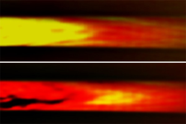
Now there’s a better way to spy on the blood in your veins. Doctors already have two techniques to monitor obstructions in blood vessels, but they both have limitations. The first, Doppler ultrasound imaging, involves irradiating tissue with ultrasound waves; the waves that reflect off flowing blood acquire a Doppler shift, which can be used to pick out blood and calculate its speed. Doppler can't distinguish flowing blood from surrounding tissue unless it's moving quickly, however, which makes minor blood vessels invisible. The second technique, photoacoustic imaging, uses an infrared laser that, when absorbed by blood, heats it. The resulting sudden expansion creates a pressure wave that can be detected outside the body. Photoacoustic imaging picks out blood vessels better, but it can't see flow in a continuous stream. In a study published today in Physical Review Letters, researchers combined the two techniques, utilizing the fact that ultrasound also has a slight heating effect; pulsed ultrasound creates periodic hot spots in blood vessels. By tracking the movement of these hot spots (shown in yellow above) using photoacoustic imaging, the team could calculate the flow rate of the blood, even when it moved slowly through small vessels like capillaries. The researchers hope their technique may aid functional brain imaging, help cancer screening and treatment monitoring, and let doctors detect atherosclerosis before a patient has a heart attack.
A new method for imaging the flow of blood has been developed by researchers in the US. By using ultrasound to thermally tag blood, along with photoacoustics to image the resulting heat flow, the new technique is considerably more sensitive than the conventional Doppler ultrasound method that is currently used. While presently at the in vitro testing stage, this technique might have a variety of clinical applications, especially in medical diagnosis...More>>
Lihong Wang, the Gene K. Beare Distinguished Professor of
Biomedical Engineering at Washington University in St. Louis, is trying to improve endoscopy by combining conventional ultrasound with a technology called photoacoustics. Ultrasound endoscopy provides high-resolution images of structures and is widely used to look for oesophageal or colorectal cancer, for example. But its contrast is low, so it does not readily distinguish between blood vessels and lymphatic vessels, or healthy and diseased soft tissue...More>>
Lihong Wang, the Gene K. Beare Distinguished Professor of Biomedical
Engineering at Washington University in St. Louis, has received a
three-year, $300,000 grant from the National Science Foundation (NSF) to
study oxygen consumption rates of individual cells using photoacoustic
microscopy, a novel imaging technology he developed that uses light and
sound to measure change...More>>
Lihong Wang, PhD, the Gene K. Beare Distinguished Professor of
Biomedical Engineering at Washington University in St. Louis, changed
his area of emphasis from electrical engineering to biomedical
engineering because he dreamed about making a difference in patients’
lives...More>>
Scientists from the University of Southern California
in Los Angeles and Washington University in St. Louis have developed a
new type of medical imaging that gives doctors a new look at live
internal organs...More>>
A collaboration between a surgeon and a biomedical
engineer led to the invention of photoacoustic endoscopy, a powerful new
tool to screen for an esophageal disorder...More>>
Photoacoustic tomography (PAT), combining optical and
ultrasonic waves via the photoacoustic effect, provides in vivo
multiscale non-ionizing functional and molecular imaging...More>>
A new imaging technique called photo-acoustic
tomography combines the properties of light and sound to give doctors a
powerful tool to detect cancer earlier than ever before. Its developers
say it can also be performed without the dangers of radiation exposure
associated with current imaging methods like x-ray and CT scans...More>>
Photoacoustic imaging is starting to be used on human
patients, and the technology could revolutionize medical imaging in
clinical practice – from early-stage cancer detection, to neurology and
label-free histology...More>>
Tracking tumors is a tricky business. Scientists have
discovered recently, however, that ‘listening’ might make it easier.
This image of a melanoma
(represented in gold) and blood vessels (in red) growing under mouse
skin, was produced by doing just that...More>>
In short, “light goes in, sound comes out,” Wang
says. Molecules absorb incoming pulses of laser light, which heat them
up a tiny, harmless amount [PDF]...More>>
A new imaging technique uses light and sound rather
than radiation and delivers a rich, photographic rendering of structures
several inches below the skin...More>>
My hope is that photoacoustic
tomography will impact both basic science research and clinical
utility,” said Lihong Wang, PhD, Gene K. Beare Distinguished
Professor of Biomedical Engineering, who details this new
imaging technology in the March 23 issue of Science...More>>
Researchers at Washington University in St Louis have
used photoacoustic tomography – a non-invasive imaging technique – to
look at how gold nanocages accumulate in the lymph nodes of rats...More>>
In vivo, label-free subwavelength-resolution
photoacoustic microscopy enables more precise measurement of optical
absorption – and, therefore, offers more information...More>>
The Optical Society (OSA) has awarded Lihong V. Wang
the C.E.K. Mees Medal for seminal contributions to photoacoustic
tomography and Monte Carlo modeling of photon transport in biological
tissues and for leadership in the international biophotonics community...More>>
A focused beam of light can trap a colloidal sphere,
causea specific neuron to fire, or deliver a lethal doseof energy to a cancerous cell. In biomedicine, focused lightcan perform nearly all the same sensing, diagnostic, and
therapeuticfunctions as targeted x rays, without inducing
harmful ionization...More>>
The ability of photoacoustic microscopy (PAM) to
produce exceptionally high spatial resolution images of cutaneous
microvascular networks could provide a vital insight into cardiovascular
diseases. That's the conclusion of researchers from Washington
University in St. Louis, MO (WUSTL)...More>>
To see life in its natural
state, researchers want—as much as possible—imaging
approaches that need no stains or labels. Photoacoustics provide
this with "rich contrast," according to Lihong Wang, Gene K. Beare
Distinguished Professor at Washington University in St. Louis, and
inventor of three-dimensional photoacoustic microscopy (see
http://bit.ly/h2EUI5)...More>>
The strong scattering of light in biological tissue
impedes the development of light-based biological imaging. Lihong Wang
explained to Nature Photonics how the use of ultrasound can aid the
deeper and tighter focusing of light in scattering media.
[Nature Photonics 5, 184 (2011), DOI: 10.1038 / nphoton. 2011.
24]...More>>
Combining ultrasonic modulation and
optical phase conjugation allows light to be tightly focused in a
scattering medium, providing benefits for studies of photophysical,
photochemical and photobiological processes [Nature Photonics 5, 135-136
(2011), DOI: 10.1038 / nphoton. 2011. 19]... More>>
Lihong Wang, PhD, the Gene K. Beare Distinguished
Professor of Biomedical Engineering at Washington University in St.
Louis, has invented a guide star for biomedical rather than celestial
imaging, a breakthrough that promises game-changing improvements in
biomedical imaging and light therapy...More>>
The team behind the research, led by Lihong Wang at
Washington University in St Louis, US, say that the high photoacoustic
sensitivity of plasmon-resonant nanostars at near-infrared wavelengths
enables the in vivo detection in rat sentinel lymph nodes and
vessels...More>>
Lihong Wang and colleagues at Washington University
in St. Louis have developed a new approach that combines time reversal
with ultrasound, whose waves scatter weakly in biological tissue, to
focus light to a controllable position...More>>
Lihong Wang from Washington University presented some
fantastic results on photoacoustic imaging in a plenary talk at the Physics of Quantum Electronics
conference, bedazzling the audience with beautiful image after beautiful
image...More>>
See it for yourself: a new breakthrough in imaging
technology using a combination of light and sound will allow health care
providers to see microscopic details inside the body...More>>
A new discovery may lead to a very early detection of
melanomas, the most serious of skin cancers that kills thousands of male
senior citizens every year...More>>
Melanoma is one of the less common types of skin
cancer but it accounts for the majority of the skin cancer deaths (about
75 percent)...Two scientists at Washington University in St. Louis have
developed technologies that together promise to solve this difficult
problem...More>>
Now Washington University professors are developing
techniques using Bell's photoacoustic effect. The new imaging technology
being developed by Lihong Wang and his colleagues will identify the
sentinel lymph node...More>>
Cover Story
<10-15> “2003 is the magic number,” says Wang, Ph.D., now the
Gene K. Beare Distinguished Professor in the Department of Biomedical
Engineering. “We published the first paper on functional imaging using
photoacoustics. That excited everybody and attracted newcomers to the
field.”...More>>
BELLINGHAM, Washington, and WASHINGTON, D.C., USA --
Lihong V. Wang and Hsin-I Wu are recipients of the 2010 Joseph W.
Goodman Book Writing Award (List
of winners) for their book Biomedical Optics: Principles
and Imaging, the Optical Society (OSA) and SPIE have announced...More>>
Photoacoustic imaging combines light and sound to
create detailed pictures of tiny structures in the body without the use
of high-energy X-ray beams, which can be damaging. Unlike traditional
radiology techniques, it also provides functional information about
tissues and cells, with the ability to show blood flow and oxygen
saturation...More>>
Researchers from the Optical Imaging Laboratory at
Washington University in St. Louis, led by Dr. Lihong Wang, have been
very active developing techniques that combine photoacoustic
microscopy and optical coherence tomography. Combining sound and
light imaging can provide several benefits including images at greater
depths with complementary contrasts...More>>
Nanotubes reveal breast cancer spread: Sentinel lymph-node biopsy is certainly less drastic than no-questions-asked underarm lymph-node dissection, but it is not without its disadvantages. Node identification typically involves injection of a gamma ray-emitting radiotracer and/or blue dye into the breast...More>>
The Sound of Light: If light passed through objects, rather than bouncing off them, people might now talk to each other on “photophones”. Alexander Graham Bell demonstrated such a device in 1880, transmitting a conversation on a beam of light...More>>
SPIE appoints Lihong Wang editor of 'Journal of Biomedical Optics': SPIE has announced the appointment of Lihong V. Wang of Washington University in St. Louis as editor of theJournal of Biomedical Optics effective 1 January 2010...More>>
Photoacoustics sheds light on brain function: Researchers at Washington University in St. Louis, MO, (WUSTL) have used photoacoustic microscopy (PAM) to obtain high-resolution in vivo images of the mouse brain in response to
differing oxygen levels...More>>
Novel technique changes lymph node biopsy, reduces radiation exposure in breast cancer patients: Ultrasonography and MRI are increasingly being used as complementary modalities for breast-cancer diagnosis...More>>
Harnessing Light and Sound for Staging of Breast Cancer: Only recently developed for imaging of tissue, photoacoustic tomography now is racing headlong toward clinical implementation...More>>
Photoacoustic Imaging Gets Dynamic: Photoacoustic imaging offers tremendous potential for both research and clinical applications because it draws on the advantages of both spectroscopy and ultrasound imaging... More>>
TAT/PAT: a new screening option? Information obtained from a new application of photoacoustic tomography (PAT) is worth its weight in gold to breast cancer patients...More>>
Researchers at the Texas A&M University have developed a new method for using the photoacoustic effect to create images. The technique allows for functional imaging of oxy and deoxyhemoglobin with an axial resolution of about 15 µm, a lateral resolution of 45 µm, and an imaging depth of 3 mm...More>>
Medical imaging researchers are
always trying to increase the
resolution and penetration of their
instruments, with the ultimate goal of
achieving the same microscopic resolution
in living tissue that can be obtained
by biopsy, thus eliminating the often
painful process of taking biopsy samples...More>>