|
early 17 embryo |
|
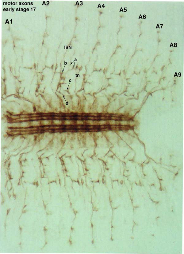
This first section (column 1) presents two brightfield images of entire dissected embryos and two photographs of the CNS. These four images are useful as references for staging embryos. Embryos are roughly staged prior to dissection using gut segmentation and CNS contraction as markers. Dissected preparations can then be more accurately staged using the 1D4 CNS staining pattern and the muscle morphologies. To stage mutant embryos, it is important to use markers such as these that are separate from the motor axons themselves, since a number of mutants cause temporal delays in motor axon branch development. The motor axons extend in 5 major branches. Three of these (ISN, SNb, and SNd) arise from the ISN root, and two (SNa and SNc) from the SN root. SNb and SNd separate from the common ISN pathway at the exit junction, and SNb and SNd then separate from each other at a nearby second junction. SNa separates from SNc at another junction point. All three of these junctions are in the ventral muscle region. The ISN and SN roots never really come together, although they approach each other closely in the exit junction region. The transverse nerve also stains with 1D4. A characteristic cell, which has been called the m cell, lies along the this nerve and is adjacent to the ISN. This is a brightfield image (10X lense) of a dissected early stage 17 embryo stained with 1D4. Anterior is to the left. Note the three distinct longitudinal bundles in the CNS, and the lack of strong commissural or cell body staining. Note also the stereotypy of the motor axons in abdominal segments A2-A7, which are the segments that are normally scored. A1 has some minor differences. a, SNa; b, SNb; c, SNc; d, SNd; tn, transverse nerve. The SNb lateral synaptic branches are clearly visible. |
|