|
diagrams of muscles |
|
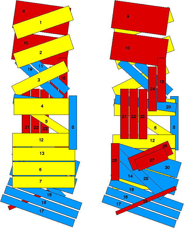
This image is based on a diagram by M. Bate (Fig. 11 on p. 1044) of the The Development of Drosophila melanogaster (Cold Spring Harbor Press, 1993). It depicts the muscle pattern in A2-A7. The muscles are divided into three arbitrary groups: yellow (interior), blue (middle, except for muscle 8), and red (exterior). The view on the left is what you would see looking through the microscope at a dissected embryo oriented in the normal manner (interior up). The view on the left is what you would see if you flattened the embryo with the exterior (epidermis) up and then focused down through the epidermis onto the muscle layer. The right-hand view, although it does not correspond to anything one sees in a normal dissection, can help in allowing one to identify the muscles that are underneath the first layer. This is often important in visualizing the trajectories of the SNb and SNa branches. For example, to look at the SNb one is often focusing down at the level of muscles 30 and 14 (see SNb images), since the SNb navigates among these muscles on its way to its targets. Similarly, to look at the SNa synapses involves focusing down to muscles 5 and 21-24 (see SNa images). To diagram of muscles and axon pathways. Click this link next. |
|
||
