|
SNc and SNd |
|
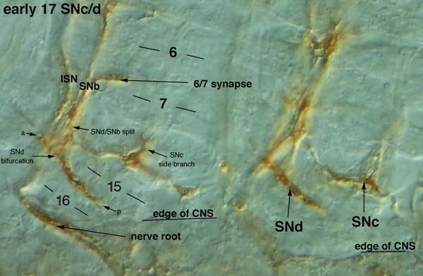
SNc and SNd are often difficult to see, because they are so close to the CNS, and they also develop late. To visualize SNc and SNd, the body walls must be pulled out so that the nerve roots do not curl up at the edge of the CNS. They can only be scored well in early 17 embryos, and because they are so short not much can be recorded about them other than whether they are present. SNc derives from the SN root; it often runs along the distal edge of muscle 15 and extends a side branch that grows dorsally. This might innervate muscles 26 and 27. SNd derives from the ISN root, and extends along the cleft between muscles 15 and 16. SNb and SNd axons both leave the ISN at the exit junction. SNd axons leave the SNb/SNd pathway distal to muscle 15 (approximate divergence point indicated as SNb/SNd split), then grow back toward the CNS and bifurcate at the 15/16 cleft. SNd in an early 17 embryo usually extends in both directions from the bifurcation point, and the anterior branch (a) sometimes crosses the ISN (as in this image). The posterior branch (p) of SNd, however, is always longer than the anterior branch. SNd is usually very straight, perhaps because its path is defined by the muscle cleft. To motor axons + PNS brightfield |
|