|
1D4-22C10 ISN |
|
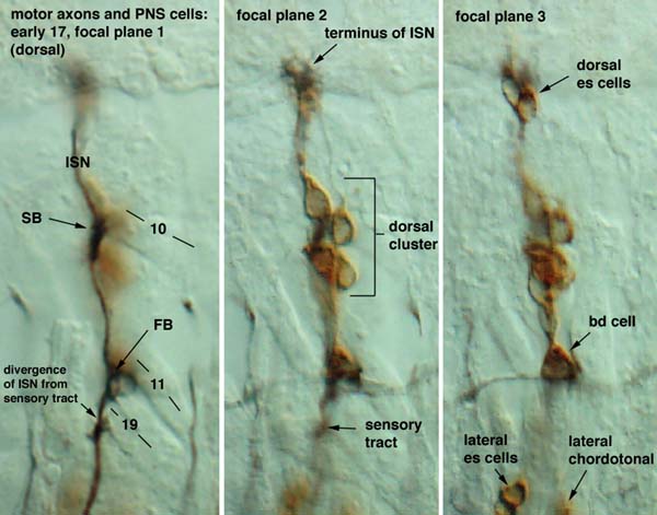
This image shows three focal planes of the dorsal region of the body wall of an early 17 embryo. In focal plane 1 the motor axons are clearly visible (black); they diverge from the sensory axons proximal to FB, around the location of the arrow. Note that the proximal section of the ISN is blackish-brown, as it contains both motor and sensory axons. The distal motor axons grow interiorly to the PNS cell bodies. In the second focal plane one sees the dorsal cluster of external sensory (es) neurons and the tract extending proximally from this cluster. In the third focal plane are the bipolar dendritic cell and the distal edges of the lateral cluster (visible in next image of the ventral region). To early 17 motor axons + PNS: ventral region To early 17 motor axons + PNS: brightfield |
|
|
|