|
motor axons + PNS-whole body wall |
|
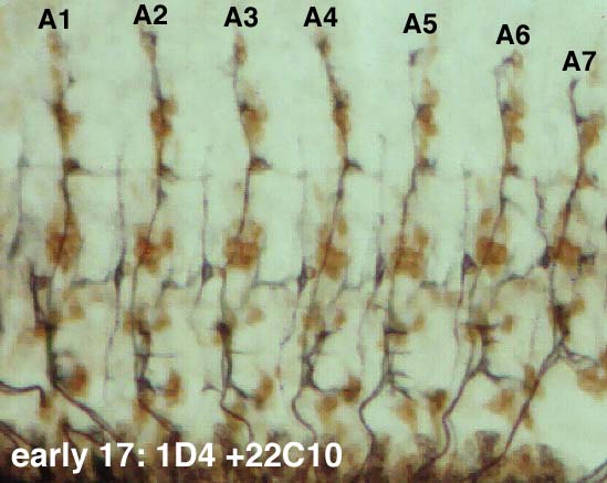
This is a brightfield image of one body wall of an embryo double stained with 1D4 (black; HRP + Ni immunohistochemistry) and MAb 22C10 (brown; HRP immunohistochemistry). 22C10 stains peripheral sensory neuron cell bodies and axons, and a subset of axons and cell bodies in the CNS (not shown). Like the motor axon pattern, the PNS pattern in A2-A7 is very stereotyped. It is described by Ghysen et al. in Roux. Arch. Dev. Biol. 195, 281-289 (1986), and in many other papers as well. To early 17 motor axons + PNS: dorsal region |
|