|
motor axons + twist: stage 14-15 |
|
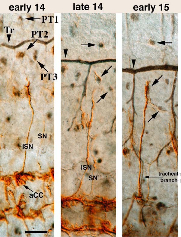
This image shows the first three panels of a 6-panel set depicting the development of the ISN pathway relative to the positions of twist-expressing PT (persistent Twist) cells (first described by M. Bate and colleagues) and the tracheal branches. These structures (black) are visualized by an anti-Twist antibody (from S. Roth) that fortuitously stains tracheal branches. PT cells are labeled in panel 1 and indicated by slanting or horizontal arrows in subsequent panels (2-6). In panel 1 the ISN has not yet reached PT2 or PT3. PT2 and PT3 are at the same position on dorsoventral axis at this stage. The staining pattern of 1D4 in the CNS at this stage is relatively simple. Only a few neurons (aCC, pCC, MP1) are stained. The cell body of aCC, the ISN pioneer, is visible. The terminal growth cones have not contacted any tracheal branch. The main tracheal trunk is indicated by Tr. Pioneer SN axons can also be seen. Panel 2: The ISN has contacted PT2. PT3 has begun migrating ventrally. Its position is now closer to the CNS than the terminal growth cones of the ISN. PT3 has apparently not yet been contacted by ISN axons. A tracheal branch is extending ventrally. The SN has almost reached the level of the m cell (adjacent to the ventral end of the tracheal side branch), and its terminus appears to be in contact with the ventral terminus of the forming tracheal branch that will eventually form the loop seen in Panel 3. The CNS 1D4 pattern is becoming more complex. Panel 3: The leading edge of the ISN terminal growth cones has passed PT2,which now lies under the most distal segment of the axon. A lateral branch has been sent out that contacts PT3. This is the FB branchpoint position. A loop of the tracheal system has been formed, and another branch (arrow) has extended ventally from the bottom of this loop and reached tracheal branches adjacent to the ISN. Note that:1) this branch was not present (or at least did not stain with anti-Twist) at the stage when the ISN was growing out; 2) the branch is not in contact with the ISN in this panel, but is adjacent to it. These facts suggest strongly that the ISN did not use the tracheal branch as a cue during its outgrowth. Rather, it seems to have grown out over the surfaces of epidermal cells and myoblasts. The SN has contacted the base of the tracheal loop and has grown posteriorly and dorsally along it. |
|