|
motor axons + twist:stage 15-16 |
|
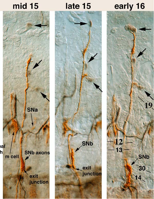
This image shows the remaining 3 panels of the 6-panel set. Panel 4: The ISN terminus is between PT2 (45 degree downward arrows) and PT1 (horizontal arrows). PT2 now lies adjacent to the ISN axons. A side branch exists to PT3 (45 degree upward arrows), but is not clearly visible here. The main tracheal trunk has been dissected away in these three panels, so only some tracheal side branches are visible. SNa has crossed over the tracheal loop. The SNb axons are at the exit junction, and can be visualized as a swelling of the common ISN pathway. Panel 5: The ISN terminus has just contacted PT1. PT2 is migrating ventrally along the ISN axons toward PT3. The FB branch at PT3 is clearly visible. The SNb has begun to extend into the ventrolateral muscle field. Panel 6: The ISN begins to form a terminal arbor around PT1. PT2 in this segment is much farther from PT3 than is the PT2 in Panel 5: this illustrates the variability in positioning of PT2. Differentiation of muscle fibers (some are numbered) is evident. The terminus of the SNb has reached the position of the 13/30 synapse. Most of the growth cone material in the SNb, however, is still located at the cleft between muscles 30 and 14. To motor axons + twist: stage 14-15 |
|