|
|
| NEMS Biosensors: Microfluidic Embedded NEMS for Medical Diagnostics | |||
| Research Lab Tour Publications People Gallery Courses Opportunities Links Contact Home
|
We are exploring new technologies for rapid (<10 minute), small volume (nL-µL), high sensitivity biodetection.
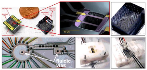
Figure 1. Assembly of microfluidics-embedded nanocantilever arrays into functional microanalysis systems. The top left panel shows front and backside views of individual 1x1 cm nanosensor chips fused front and back with microfluidic systems. The top central panel is a scanning electron microscope (SEM) image of two of the MEMS sensors at the heart of the device. The top right and bottom left panels are progressive magnifications of the microfluidic delivery system on the backside of the chip, showing the 70 µm x 70 µm fluidic vias through the chip (comprising a total volume of 1.5 µL) coupled to 16 analyte delivery channels, a peristaltic sample recirculation pump, and a valved sample exhaust port. The total volume including delivery channel, circulation loop, exhaust port, and 2 fluidic vias is of order 4 µL. The two panels on the bottom right show a top/bottom view of a current-generation 2x3 cm chip mount which provides up to 40 fluidic input/output lines and electrical connections for up to 20 nanocantilevers. The ability to perform multiparameter testing, on blood volumes of ~10µL or less in less than 20 minutes could revolutionalize medical diagnostic lab work, allowing for point of care diagnosis for a tremendous range of disorders and pathogens. To achieve this will require new technologies. We are exploring novel geometries for MEMS and NEMS-based concentration sensing. Figure 1 shows a silicon MEMS cantilever embedded in microfluidics. Nanoscale devices have great promise for enhanced sensitivity. However, for devices immersed in fluid this sensitivity is frequently compromised by heavy damping. We are exploring novel geometries to achieve the greatest possible sensitivity with minimal damping. Critical to the success of these efforts will be the large scale integration of arrays of devices towards a nanosystem. This effort therefore ties in closely with other efforts in the group in this area. Chemical recognition is a critical component to achieving specificity across almost all biodetection platforms. With this in mind we are pursuing both traditional and novel forms of device functionalization. Most of what we have learned from biology comes from studying large ensembles of cells. With these techniques we will have the ability to monitor a matrix of cells—with resolution at the individual cell level. This has the potential to open entire fields of research, with possibilities still waiting to be discovered. Personnel References
|
||
| | local users |
|||